 뒤로
Integumentary System: Structure, Function, and Accessory Organs
스터디 가이드 - 스마트 노트
 자료에 맞춘 맞춤형 노트, 핵심 정의, 예시, 맥락을 확장해 제공합니다.
The Integumentary System
Overview and General Functions
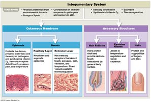
The integumentary system is the largest organ system in the human body, comprising approximately 16% of body weight. It consists of the skin (cutaneous membrane) and its accessory structures. The system provides protection, regulates temperature, synthesizes nutrients, and enables sensory reception.
Protection: Shields underlying tissues from mechanical, chemical, and microbial damage.
Temperature Regulation: Maintains body temperature through insulation and sweat evaporation.
Synthesis and Storage: Epidermis synthesizes vitamin D3; dermis stores lipids.
Sensory Reception: Detects pain, touch, pressure, and temperature.
Excretion and Secretion: Glands excrete salts, water, and organic wastes; mammary glands secrete milk.
Components of the Integument
The integument consists of two main components:
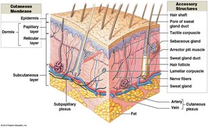
Cutaneous Membrane: Composed of the epidermis (superficial layer) and dermis (deeper connective tissue layer).
Accessory Structures: Originate in the dermis and extend through the epidermis, including hair, nails, and exocrine glands.
Structure of the Skin
Epidermis
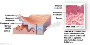
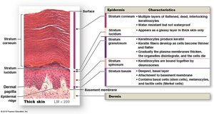
The epidermis is an avascular, stratified squamous epithelium. It is divided into layers (strata) that differ between thick and thin skin.
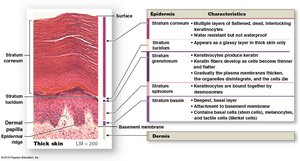
Thick Skin: Found on palms and soles; contains five strata.
Thin Skin: Found elsewhere; contains four strata.
Strata of the Epidermis
Stratum Basale: Deepest layer; attached to basement membrane via hemidesmosomes. Contains stem cells (germinative cells), melanocytes, and Merkel cells. Forms epidermal ridges (fingerprints).
Stratum Spinosum: "Spiny layer"; 8–10 layers of keratinocytes. Contains dendritic cells for immune response.
Stratum Granulosum: "Grainy layer"; 3–5 layers. Keratinization begins here; cells die and become waterproof.
Stratum Lucidum: Found only in thick skin; flat, keratin-filled cells without organelles.
Stratum Corneum: Most superficial; 15–30 layers of dead, keratinized cells. Cells are shed and replaced every 2 weeks.
Dermis
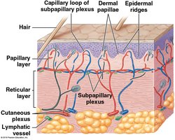
The dermis lies beneath the epidermis and is divided into two layers:
Papillary Layer: Superficial; made of areolar connective tissue. Contains dermal papillae, capillary loops, and sensory receptors (Meissner’s corpuscles, free nerve endings).
Reticular Layer: Deep; dense, irregular connective tissue with collagen and elastic fibers. Contains larger blood vessels, lymphatic vessels, and nerves (lamellated corpuscles).

Hypodermis (Subcutaneous Layer)
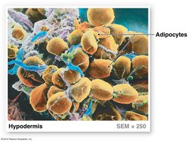
The hypodermis is not part of the integument but stabilizes the skin to underlying tissues. It consists of areolar tissue and adipose cells, serving as a site for subcutaneous injections.
Function: Provides insulation, energy storage, and protection.
Cells of the Epidermis
Keratinocytes
Most abundant epidermal cells.
Produce keratin, a tough, fibrous protein that provides waterproofing.
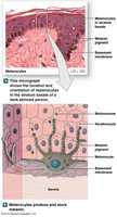
Melanocytes
Located in the stratum basale.
Produce melanin, which protects against UV radiation and contributes to skin color.
Skin color differences are due to the rate of melanin synthesis, not the number of melanocytes.
Merkel Cells
Found in areas lacking hair (palms, soles).
Function as tactile receptors for light touch.
Dendritic Cells
Located in the stratum spinosum.
Activate immune responses against pathogens and superficial skin cancers.
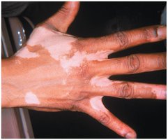
Skin Color and Disorders
Factors Affecting Skin Color
Skin color is determined by the presence of melanin, carotene, and dermal blood flow.
Melanin: Brown pigment produced by melanocytes; protects against UV light.
Carotene: Orange-yellow pigment from diet; source of vitamin A.
Dermal Blood Flow: Oxygenated blood gives a reddish color; vasodilation causes flushing, vasoconstriction causes paling.
Common Skin Disorders
Albinism: Genetic condition; melanocytes present but unable to synthesize melanin.
Cyanosis: Bluish coloration due to diminished oxygen in blood.
Jaundice: Yellowish color due to liver dysfunction.
Vitiligo: Loss of color due to loss of melanocytes.
Effects of Sunlight and Vitamin D3 Synthesis
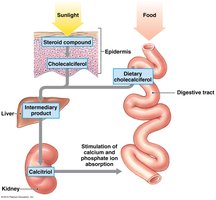
Vitamin D3 Production
Exposure to sunlight triggers the synthesis of cholecalciferol (vitamin D3) in epidermal cells. Vitamin D3 is converted to calcitriol in the kidneys, which increases calcium and phosphorus absorption in the small intestine.
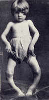
Deficiency: Leads to rickets, characterized by weak and flexible bones.
Accessory Structures of the Integument
Hair and Hair Follicles
Hair is a nonliving structure produced by hair follicles located deep in the dermis and hypodermis.
Functions: Protects and insulates the scalp, prevents entry of foreign particles, aids in sensory perception, and expresses emotional state (goosebumps).
Structure: Hair root (attached, not visible), hair shaft (visible).
Hair Matrix: Layer of dividing basal cells that produce hair; cells become keratinized and die as they move upward.
Hair Shaft Layers: Medulla (soft keratin), cortex (hard keratin), cuticle (hard keratin).
Glands
Sebaceous Glands: Near hair roots; secrete oily sebum via holocrine secretion to lubricate hair and skin, inhibit bacterial growth. Blockage leads to acne.
Sebaceous Follicles: Secrete sebum directly onto skin (face, back, chest, nipples, genitalia).
Apocrine Sweat Glands: Located in armpits, nipples, pubic areas; produce sticky, cloudy secretions into hair follicles. Odor caused by bacterial breakdown.
Merocrine (Eccrine) Sweat Glands: Secrete directly onto skin surface; widely distributed, especially on palms and soles. Function in thermoregulation.
Modified Sweat Glands: Mammary glands (milk production), ceruminous glands (earwax production).
Nails
Nails protect the tips of fingers and toes.
Nail Body: Visible part; mass of dead, keratinized cells.
Nail Bed: Lies beneath the nail body.
Nail Root: Site of nail production, deep in epidermal fold.
Lunula: Pale crescent at the base of the nail.
Eponychium (cuticle): Proximal end, covers root.
Hyponychium: Skin beneath distal free edge.
Repair and Regeneration of Skin
Four Stages of Tissue Repair
Inflammatory Phase: Bleeding and mast cell activation initiate inflammation.
Migratory Phase: Scab forms; basale cells migrate to isolate area; phagocytic cells clean debris; granulation tissue forms.
Proliferation Phase: Epidermal cells cover connective tissue meshwork; clot dissolves; fibroblasts secrete proteins.
Scarring Phase: Scab is shed; epidermis is complete; scar tissue forms and gradually elevates.
Summary Table: Layers and Functions of the Integument
Layer | Structure | Function |
|---|---|---|
Epidermis | Stratum basale, spinosum, granulosum, lucidum (thick skin), corneum | Protection, waterproofing, vitamin D3 synthesis |
Dermis | Papillary layer (areolar CT), reticular layer (dense irregular CT) | Support, nourishment, flexibility, sensory reception |
Hypodermis | Areolar tissue, adipose cells | Insulation, energy storage, protection |
Accessory Structures | Hair, glands, nails | Protection, lubrication, thermoregulation, sensory input |
Additional info:
Keratinization is the process by which cells become filled with keratin and die, forming a protective, waterproof layer.
The average skin cell life cycle is about 21 days.
Dermal circulation is crucial for thermoregulation and skin color changes.
