 뒤로
Integumentary System Study Guide – Anatomy & Physiology
스터디 가이드 - 스마트 노트
 자료에 맞춘 맞춤형 노트, 핵심 정의, 예시, 맥락을 확장해 제공합니다.
Q1. Describe the components of the integumentary system and how they contribute to homeostasis.
Background
Topic: Integumentary System Overview
This question tests your understanding of the main parts of the integumentary system (skin, hair, nails, glands) and their roles in maintaining internal balance (homeostasis).
Key Terms:
Integumentary system: Skin, hair, nails, glands
Homeostasis: The body's ability to maintain stable internal conditions
Step-by-Step Guidance
List the main components: skin, hair, nails, and glands.
Describe how each component helps protect the body (e.g., skin as a barrier, hair for insulation).
Explain how the skin regulates temperature and prevents dehydration.
Discuss the role of glands (sweat and sebaceous) in homeostasis.
Consider how the integumentary system interacts with other systems to maintain balance.
Try solving on your own before revealing the answer!
Final Answer:
The integumentary system includes the skin, hair, nails, and glands. The skin acts as a barrier against pathogens and dehydration, regulates temperature through sweat glands, and produces vitamin D. Hair provides insulation and protection, nails protect the fingertips, and glands (sebaceous and sweat) help with lubrication and temperature regulation. Together, these components maintain homeostasis by protecting the body, regulating temperature, and preventing water loss.
Q2. Describe the skin, including the functions of its cell types and layers.
Background
Topic: Skin Structure and Function
This question focuses on the anatomy of the skin, its layers (epidermis, dermis, hypodermis), and the functions of different cell types found within these layers.
Key Terms:
Epidermis: Outer layer, mainly keratinocytes
Dermis: Middle layer, contains connective tissue, blood vessels
Hypodermis: Deepest layer, mostly fat
Cell types: Keratinocytes, melanocytes, Langerhans cells, Merkel cells
Step-by-Step Guidance
Identify the three main layers: epidermis, dermis, hypodermis.
List the cell types found in the epidermis and their functions (e.g., keratinocytes produce keratin).
Describe the role of melanocytes, Langerhans cells, and Merkel cells.
Explain how the dermis supports the epidermis and contains structures like blood vessels and nerves.
Discuss the hypodermis and its function in insulation and energy storage.
Try solving on your own before revealing the answer!
Final Answer:
The skin consists of three layers: the epidermis (with keratinocytes for protection, melanocytes for pigment, Langerhans cells for immunity, and Merkel cells for sensation), the dermis (providing structural support, blood supply, and sensory functions), and the hypodermis (mainly fat for insulation and energy storage). Each cell type and layer contributes to the skin's protective, sensory, and regulatory functions.
Q3. What are the functions of the integumentary system?
Background
Topic: Functions of the Integumentary System
This question tests your knowledge of the various roles the integumentary system plays in the body.
Key Terms:
Protection
Thermoregulation
Sensation
Vitamin D synthesis
Excretion
Step-by-Step Guidance
List the main functions: protection, thermoregulation, sensation, vitamin D synthesis, excretion.
Explain how the skin acts as a barrier against pathogens and injury.
Describe how sweat glands and blood vessels help regulate body temperature.
Discuss the sensory receptors in the skin and their role in detecting stimuli.
Consider how the skin helps synthesize vitamin D and excretes waste products.
Try solving on your own before revealing the answer!
Final Answer:
The integumentary system protects the body from physical, chemical, and biological harm, regulates temperature, provides sensory information, synthesizes vitamin D, and excretes waste through sweat. These functions are essential for maintaining health and homeostasis.
Q4. The major layers of the integument are the epidermis and the dermis. What type of tissue make up each of these layers?
Background
Topic: Tissue Types in Skin Layers
This question tests your understanding of the histology of the skin, specifically the types of tissue found in the epidermis and dermis.
Key Terms:
Epidermis: Stratified squamous epithelium
Dermis: Connective tissue (dense irregular)
Step-by-Step Guidance
Identify the tissue type in the epidermis (epithelial tissue).
Describe the tissue type in the dermis (connective tissue).
Explain the structural differences between these tissues.
Discuss how these tissue types contribute to the skin's function.
Try solving on your own before revealing the answer!
Final Answer:
The epidermis is made of stratified squamous epithelial tissue, which provides protection. The dermis is composed of dense irregular connective tissue, which gives strength and elasticity to the skin.
Q5. What are the four cell types that make up the epidermis? What is the function of these cells?
Background
Topic: Epidermal Cell Types
This question tests your knowledge of the cellular composition of the epidermis and the specific functions of each cell type.
Key Terms:
Keratinocytes: Produce keratin
Melanocytes: Produce melanin
Langerhans cells: Immune defense
Merkel cells: Sensory reception
Step-by-Step Guidance
List the four cell types: keratinocytes, melanocytes, Langerhans cells, Merkel cells.
Describe the function of keratinocytes in producing keratin for protection.
Explain the role of melanocytes in pigment production and UV protection.
Discuss the immune function of Langerhans cells.
Describe the sensory function of Merkel cells.
Try solving on your own before revealing the answer!
Final Answer:
The four cell types in the epidermis are keratinocytes (protection), melanocytes (pigment and UV protection), Langerhans cells (immune defense), and Merkel cells (touch sensation).
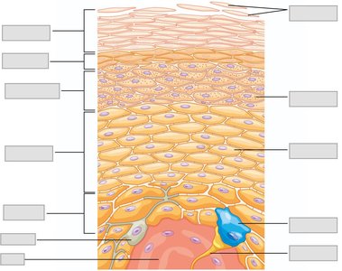
Q6. What are the layers of the epidermis? What happens at each of these layers? Identify these layers on an image.
Background
Topic: Epidermal Layers
This question tests your knowledge of the five layers of the epidermis and the processes occurring in each layer.
Key Terms:
Stratum basale
Stratum spinosum
Stratum granulosum
Stratum lucidum (only in thick skin)
Stratum corneum
Step-by-Step Guidance
List the five layers from deep to superficial.
Describe the main function or process in each layer (e.g., cell division in stratum basale).
Explain the presence of stratum lucidum in thick skin only.
Discuss the process of keratinization as cells move upward.
Use the provided image to identify and label each layer visually.

Try solving on your own before revealing the answer!
Final Answer:
The five layers of the epidermis are (from deep to superficial): stratum basale (cell division), stratum spinosum (cell strength), stratum granulosum (keratinization), stratum lucidum (only in thick skin), and stratum corneum (dead, keratinized cells). The image shows these layers and their arrangement.
Q7. What is keratinization? Describe what happens in this process and why it is important.
Background
Topic: Keratinization Process
This question tests your understanding of how epidermal cells change as they move toward the skin surface and why this process is vital for protection.
Key Terms:
Keratinization: Formation of keratin in cells
Keratin: Protective protein
Step-by-Step Guidance
Describe how keratinocytes originate in the stratum basale.
Explain how these cells move upward through the epidermal layers.
Discuss the accumulation of keratin and loss of organelles.
Explain why keratinization is important for waterproofing and protection.
Try solving on your own before revealing the answer!
Final Answer:
Keratinization is the process by which keratinocytes produce and accumulate keratin as they move from the stratum basale to the stratum corneum. This process results in the formation of a tough, protective, and waterproof layer that prevents dehydration and protects against environmental damage.
Q8. What are the two regions of the dermis? What happens in each of the regions?
Background
Topic: Dermal Structure
This question tests your knowledge of the dermis, its subdivisions, and the functions of each region.
Key Terms:
Papillary region: Superficial, loose connective tissue
Reticular region: Deep, dense connective tissue
Step-by-Step Guidance
Identify the papillary and reticular regions of the dermis.
Describe the structure and function of the papillary region (e.g., contains capillaries, sensory receptors).
Explain the structure and function of the reticular region (e.g., provides strength, contains glands and hair follicles).
Discuss how these regions support the epidermis and overall skin function.
Try solving on your own before revealing the answer!
Final Answer:
The dermis has two regions: the papillary region (superficial, contains capillaries and sensory receptors) and the reticular region (deep, provides strength and houses glands and hair follicles). Each region supports the skin's structure and function.
Q9. Label the image and be able to identify a similar image on the exam.
Background
Topic: Skin Anatomy Identification
This question tests your ability to visually identify and label the layers and structures of the skin in a diagram.
Key Terms:
Epidermal layers
Dermal regions
Accessory structures
Step-by-Step Guidance
Review the diagram and identify the main layers: epidermis, dermis, hypodermis.
Locate and label the five epidermal layers.
Identify accessory structures such as glands, hair follicles, and nerves.
Practice labeling similar diagrams to reinforce your knowledge.
Try solving on your own before revealing the answer!
Final Answer:
The image should be labeled with the five epidermal layers, dermal regions, and accessory structures such as glands and nerves. Practice identifying these features for exam readiness.
Q10. How would a tattoo be administered – through what layers of the skin and why is it permanent?
Background
Topic: Tattoo Administration and Permanence
This question tests your understanding of skin anatomy and why tattoos remain permanent.
Key Terms:
Dermis: Layer where tattoo ink is deposited
Permanence: Ink remains because dermal cells do not shed
Step-by-Step Guidance
Describe the process of tattooing and the layers involved.
Explain why the ink must reach the dermis for permanence.
Discuss why tattoos are not removed by normal skin shedding.
Consider the role of immune cells in tattoo fading over time.
Try solving on your own before revealing the answer!
Final Answer:
A tattoo is administered by injecting ink into the dermis, which is below the epidermis. The ink remains permanent because the dermal cells do not shed like epidermal cells, so the pigment stays in place.
Q11. What are epidermal ridges?
Background
Topic: Epidermal Ridges
This question tests your knowledge of the structural features of the skin that contribute to grip and identification.
Key Terms:
Epidermal ridges: Fingerprints
Dermal papillae: Underlying structures
Step-by-Step Guidance
Define epidermal ridges and their location.
Explain how they are formed by the interaction between the epidermis and dermis.
Discuss their function in increasing grip and sensitivity.
Consider their role in identification (fingerprints).
Try solving on your own before revealing the answer!
Final Answer:
Epidermal ridges are patterns on the skin surface (fingerprints) formed by the interaction of the epidermis and dermal papillae. They increase grip and sensitivity and are unique to each individual.
Q12. What are the colors / pigments of skin? How can we use this knowledge clinically? What are some pathologies associated with skin color?
Background
Topic: Skin Pigments and Clinical Relevance
This question tests your understanding of skin color, its pigments, and how changes in color can indicate health issues.
Key Terms:
Melanin: Brown/black pigment
Carotene: Yellow/orange pigment
Hemoglobin: Red pigment
Pathologies: Jaundice, cyanosis, albinism
Step-by-Step Guidance
List the main skin pigments: melanin, carotene, hemoglobin.
Describe how each pigment contributes to skin color.
Explain how changes in skin color can indicate clinical conditions (e.g., jaundice, cyanosis).
Discuss pathologies associated with abnormal skin color.
Try solving on your own before revealing the answer!
Final Answer:
Skin color is determined by melanin, carotene, and hemoglobin. Clinically, changes in skin color can indicate conditions like jaundice (yellow), cyanosis (blue), or albinism (lack of pigment). These observations help diagnose underlying health issues.
Q13. What are the accessory structures of skin?
Background
Topic: Skin Accessory Structures
This question tests your knowledge of the structures associated with the skin that contribute to its function.
Key Terms:
Hair
Nails
Glands (sebaceous, sweat)
Step-by-Step Guidance
List the accessory structures: hair, nails, glands.
Describe the function of each structure.
Explain how these structures support the skin's protective and regulatory roles.
Try solving on your own before revealing the answer!
Final Answer:
Accessory structures of the skin include hair (protection and sensation), nails (protection), and glands (sebaceous for lubrication, sweat for thermoregulation).
Q14. What is the structure of hair? What is the function of hair? How does hair grow?
Background
Topic: Hair Structure and Growth
This question tests your understanding of the anatomy of hair, its functions, and the process of hair growth.
Key Terms:
Hair follicle
Hair shaft
Growth cycle: Anagen, catagen, telogen
Step-by-Step Guidance
Describe the structure of hair (follicle, shaft, root).
Explain the function of hair (protection, sensation).
Discuss the hair growth cycle (anagen, catagen, telogen).
Explain how hair grows from the follicle.
Try solving on your own before revealing the answer!
Final Answer:
Hair consists of a shaft, root, and follicle. Its functions include protection and sensation. Hair grows in cycles (anagen, catagen, telogen) from the follicle, where cells divide and push older cells upward.
Q15. What are the glands in skin? What do each of these glands produce? Where are they located?
Background
Topic: Skin Glands
This question tests your knowledge of the types of glands in the skin, their products, and their locations.
Key Terms:
Sebaceous glands: Produce sebum
Sudoriferous glands: Produce sweat
Apocrine and eccrine glands
Step-by-Step Guidance
List the main types of skin glands: sebaceous, eccrine, apocrine.
Describe what each gland produces (sebum, sweat).
Explain where each gland is located in the skin.
Discuss the function of each gland's secretion.
Try solving on your own before revealing the answer!
Final Answer:
Sebaceous glands produce sebum and are found near hair follicles. Eccrine glands produce watery sweat and are distributed throughout the skin. Apocrine glands produce thicker sweat and are located in specific areas like the armpits.
Q16. What is the structure of nails? How do nails grow?
Background
Topic: Nail Structure and Growth
This question tests your understanding of nail anatomy and the process of nail growth.
Key Terms:
Nail plate
Nail bed
Nail matrix
Step-by-Step Guidance
Describe the structure of the nail (plate, bed, matrix).
Explain how the nail matrix produces new nail cells.
Discuss how nails grow outward from the matrix.
Consider the function of nails in protection and manipulation.
Try solving on your own before revealing the answer!
Final Answer:
Nails consist of a plate, bed, and matrix. Growth occurs as new cells are produced in the matrix and pushed outward, forming the hard nail plate. Nails protect fingertips and aid in manipulation.
Q17. What is the difference between thin and thick skin? Where is each found?
Background
Topic: Thin vs. Thick Skin
This question tests your knowledge of the structural differences between thin and thick skin and their locations.
Key Terms:
Thin skin: Covers most of the body
Thick skin: Palms, soles
Stratum lucidum: Present in thick skin
Step-by-Step Guidance
Define thin and thick skin.
Describe the structural differences (e.g., presence of stratum lucidum in thick skin).
List locations where each type is found.
Discuss functional differences (e.g., protection, grip).
Try solving on your own before revealing the answer!
Final Answer:
Thin skin covers most of the body and lacks the stratum lucidum. Thick skin is found on the palms and soles, has an extra layer (stratum lucidum), and provides greater protection and grip.
Q18. What are the different types of glands? Where are they located, what do they secrete, and how does that secretion affect the gland/cell itself?
Background
Topic: Types of Skin Glands
This question tests your knowledge of gland types, their locations, secretions, and effects.
Key Terms:
Merocrine (eccrine) glands
Apocrine glands
Holocrine glands
Step-by-Step Guidance
List the types of glands: merocrine, apocrine, holocrine.
Describe their locations and what they secrete.
Explain how secretion affects the gland or cell (e.g., cell remains intact or is destroyed).
Discuss the physiological role of each secretion.
Try solving on your own before revealing the answer!
Final Answer:
Merocrine glands secrete sweat and remain intact, apocrine glands release part of the cell with secretion, and holocrine glands release entire cells (sebaceous). Locations and effects vary by gland type.
Q19. What are the different types of burns? Explain the characteristics of each.
Background
Topic: Burn Types and Characteristics
This question tests your knowledge of burn classification and the features of each type.
Key Terms:
First-degree burn: Epidermis only
Second-degree burn: Epidermis and dermis
Third-degree burn: Full thickness
Step-by-Step Guidance
List the three types of burns: first, second, third degree.
Describe the layers affected by each burn.
Explain the symptoms and healing process for each type.
Discuss potential complications.
Try solving on your own before revealing the answer!
Final Answer:
First-degree burns affect only the epidermis (redness, pain), second-degree burns affect the epidermis and dermis (blisters, more pain), and third-degree burns destroy all skin layers (no pain, risk of infection).
Q20. What is a melanoma? How do we diagnose it?
Background
Topic: Melanoma and Diagnosis
This question tests your knowledge of skin cancer, specifically melanoma, and diagnostic criteria.
Key Terms:
Melanoma: Cancer of melanocytes
ABCDE criteria: Asymmetry, Border, Color, Diameter, Evolving
Step-by-Step Guidance
Define melanoma and its origin in melanocytes.
Describe the ABCDE criteria for diagnosis.
Explain the importance of early detection.
Discuss diagnostic methods (visual exam, biopsy).
Try solving on your own before revealing the answer!
Final Answer:
Melanoma is a cancer of melanocytes. Diagnosis uses the ABCDE criteria: Asymmetry, Border irregularity, Color variation, Diameter >6mm, and Evolving changes. Early detection is critical for treatment.
Q21. What is the benefit of the skin being multi-layered?
Background
Topic: Multi-layered Skin Benefits
This question tests your understanding of how the structure of the skin contributes to its function.
Key Terms:
Protection
Regeneration
Specialization
Step-by-Step Guidance
Explain how multiple layers provide protection against injury and pathogens.
Discuss the regenerative capacity of the basal layer.
Describe how different layers specialize in various functions (e.g., sensation, strength).
Try solving on your own before revealing the answer!
Final Answer:
Multi-layered skin offers enhanced protection, allows for regeneration, and enables specialized functions in different layers, making it highly effective as a barrier and sensory organ.
Q22. Explain why skin cancer is so common (relate to external environment and the skin cells/tissue type).
Background
Topic: Skin Cancer Prevalence
This question tests your understanding of the factors contributing to the high incidence of skin cancer.
Key Terms:
UV exposure
Cell division
Mutation risk
Step-by-Step Guidance
Explain how the skin is exposed to environmental factors like UV radiation.
Discuss the high rate of cell division in the epidermis.
Describe how mutations can occur and lead to cancer.
Consider the tissue type and its vulnerability.
Try solving on your own before revealing the answer!
Final Answer:
Skin cancer is common because the skin is constantly exposed to UV radiation, and epidermal cells divide rapidly, increasing the risk of mutations. The tissue type is vulnerable to environmental damage, leading to a higher incidence of cancer.
Q23. How is the skin involved in thermoregulation? What are the components of the skin involved and what do they do to help with thermoregulation?
Background
Topic: Skin and Thermoregulation
This question tests your understanding of how the skin helps regulate body temperature and maintain homeostasis.
Key Terms:
Sweat glands
Blood vessels
Evaporation
Step-by-Step Guidance
Describe the role of sweat glands in cooling the body.
Explain how blood vessels in the dermis dilate or constrict to regulate heat loss.
Discuss the process of evaporation and its effect on temperature.
Consider how these mechanisms maintain body temperature homeostasis.
Try solving on your own before revealing the answer!
Final Answer:
The skin regulates temperature through sweat glands (evaporation cools the body) and blood vessels (dilation increases heat loss, constriction conserves heat). These mechanisms help maintain body temperature homeostasis.
