 뒤로
Joints and Muscle Tissue: Study Guide for Anatomy & Physiology
스터디 가이드 - 스마트 노트
 자료에 맞춘 맞춤형 노트, 핵심 정의, 예시, 맥락을 확장해 제공합니다.
Joints (Articulations)
Definition and Function of Articulations
Articulations, also known as joints, are the sites where two or more bones meet. They allow for movement of the body, hold bones together, and sometimes provide protection.
Immovable joints (e.g., skull sutures) offer protection.
Movable joints enable a wide range of body movements.
Classification of Joints by Structure
Joints are classified structurally based on the material binding the bones and whether a joint cavity is present.
Fibrous Joints: Bones connected by collagen fibers; little to no movement.
Sutures: Short fibers, synarthrosis (no movement), found in the skull; closed suture is called synostoses.
Syndesmosis: Longer fibers, amphiarthrosis (slight movement), found in ligaments (e.g., tibia/fibula, radius/ulna).
Gomphosis: Periodontal ligament, synarthrosis, found in tooth roots.
Cartilaginous Joints: Bones joined by cartilage; lack a joint cavity.
Synchondrosis: Hyaline cartilage, found in epiphyseal plate and costal cartilage of 1st rib; synarthrosis.
Symphysis: Fibrocartilage (sometimes hyaline), found in intervertebral discs and pubic symphysis; amphiarthrosis.
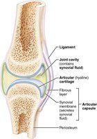
Synovial Joints: Bones connected by a fluid-filled cavity, articular cartilage, and an articular capsule; all are diarthrotic (freely movable).
Types: Plane, Condylar, Hinge, Saddle, Pivot, Ball-and-socket.
Classification of Joints by Function
Synarthroses: Immobile joints
Amphiarthroses: Slightly mobile joints
Diarthroses: Freely movable joints
General Structure of Cartilaginous Joints
Cartilaginous joints are sandwiched between bones (e.g., pubic symphysis, intervertebral discs) or at the end of a long bone and the sternum. They lack a joint cavity.
Symphysis: Example - pubic symphysis, intervertebral disc
Synchondrosis: Example - costal cartilage in sternum, epiphyseal plate
Structural Characteristics of Synovial Joints
Synovial joints are characterized by six general features:
Articular cartilage: Prevents crushing of bone ends
Articular capsule: Dense irregular connective tissue; fibrous layer strengthens joint, synovial membrane produces synovial fluid
Joint cavity: Unique to synovial joints; space filled with synovial fluid
Synovial fluid: Lubricates joint surfaces, removes debris and microbes
Reinforcing ligaments: Strengthen joints
Nerves and blood vessels: Monitor pain, joint position, and provide nutrients
Bursae and Tendon Sheaths
Bursae: Synovial membrane and fluid; reduce friction between ligaments, muscles, skin, tendons, and bones.
Tendon sheaths: Similar to bursae but wrap around tendons (e.g., wrist) to reduce friction.
Factors Stabilizing Synovial Joints
Shape of articular surfaces
Muscle tone
Number and positioning of ligaments
Common Body Movements
Gliding: Flat surfaces slide over one another
Angular movements:
Flexion: Decreasing angle between bones
Extension: Straightening a limb
Abduction: Movement away from midline
Adduction: Movement toward midline
Circumduction: Distal end moves in a circle
Rotation:
Medial rotation: Toward median plane
Lateral rotation: Toward lateral plane
Types of Synovial Joints and Examples
Plane joint: Intercarpal joints, intervertebral surfaces (gliding)
Hinge joint: Elbow, interphalangeal joints (flexion/extension)
Pivot joint: Proximal radioulnar, atlantoaxial joint (rotation)
Condylar joint: Knuckle, wrist (flexion/extension, adduction/abduction)
Saddle joint: Carpometacarpal of thumb (adduction/abduction, flexion/extension)
Ball-and-socket joint: Shoulder, hip (flexion/extension, adduction/abduction, rotation)
Major Joints: Structure, Movements, Stability, and Injury
Shoulder: Head of humerus and glenoid cavity; ball-and-socket; diarthrotic; stabilized by rotator cuffs; prone to dislocation.
Elbow: Humerus, radius, ulna; hinge; diarthrotic; stabilized by collateral ligaments; hyperextension injury.
Hip: Head of femur and acetabulum; ball-and-socket; diarthrotic; stabilized by acetabular labrum; dislocation possible.
TMJ: Mandible and temporal bone; gliding/hinge; diarthrotic; stabilized by articular disc and ligaments; easily dislocated.
Knee: Femur, tibia, patella; hinge/gliding; diarthrotic; stabilized by menisci; injuries to collateral/cruciate ligaments and cartilage.
Common Joint Injuries
Cartilage tears: Joint lock/bind, poor self-repair, may require surgery; can lead to osteoarthritis.
Sprains: Ligaments stretched/torn; poor repair; may require grafts or healing time.
Dislocations: Bones forced out of alignment; accompanied by sprains, inflammation, difficulty moving; treated by reduction.
3 C's of Knee Injury: Collateral ligaments, cruciate ligaments, cartilages (menisci); "Unhappy triad" = ruptured ACL, ruptured tibial collateral ligament, torn meniscus.
Types of Arthritis
Bursitis: Inflammation of bursae; caused by blow/friction; treated with rest, ice, anti-inflammatory drugs.
Tendonitis: Inflammation of tendons; caused by overuse; similar symptoms/treatment as bursitis.
Osteoarthritis: Degenerative, irreversible; cartilage breakdown; bone spurs; stiffness, crepitus; treated with exercise, pain relievers.
Rheumatoid arthritis: Autoimmune; joint pain, swelling, anemia, osteoporosis; inflammation of synovial membrane; treated with steroids, immune suppressants.
Gouty arthritis: Uric acid crystals in joints; affects big toe; untreated leads to joint immobilization; treated with medication, hydration, diet changes.
Lyme Disease
Caused by bacteria from tick bites; symptoms include rash, flu-like symptoms, joint pain.
Treated with antibiotics.
Less common on West Coast due to Western Fence lizard's blood killing the bacteria.
Muscles & Muscle Tissue
Types of Muscle Tissue
Skeletal muscle: Attached to skeleton; longest fibers; striated; multinucleated; voluntary; powerful but tires easily.
Cardiac muscle: Only in heart; striated; involuntary.
Smooth muscle: Walls of hollow organs; no striations; elongated cells; involuntary; moves fluids.
Muscle Terminology
Myo and mys: Muscle
Sarco: Flesh
Characteristics of Muscle Tissue
Excitability: Ability to respond to stimuli
Contractibility: Ability to shorten forcibly
Extensibility: Ability to stretch
Elasticity: Ability to return to original shape
Functions of Muscle Tissue
Produce movement
Maintain posture
Stabilize joints
Generate heat
Gross Structure of Skeletal Muscle
Made of nerves, arteries, veins, connective tissue sheaths, tendons, aponeuroses
Epimysium: Covers whole muscle
Perimysium: Covers fascicle
Endomysium: Covers muscle fiber
Microscopic Structure of Muscle Fibers
Nuclei
Sarcolemma: Plasma membrane
Sarcoplasm: Cytoplasm
T-Tubules: Allow action potential to travel
Sarcoplasmic reticulum: Stores/releases Ca2+
Actin, troponin, tropomyosin: Thin filaments
Myosin: Thick filaments
Z disc, A band, I band, H band, M line
Functional Roles in Muscle Fibers
Myofibrils: Contain sarcomeres, basis for contraction
Myofilaments: Proteins for contraction
T-tubules: Conduct action potential
Sarcoplasmic reticulum: Releases Ca2+
Sliding Filament Model of Muscle Contraction
Thin filaments slide past thick filaments, causing overlap. The I band and Z disc shorten, H zone disappears, but filament length remains unchanged.
Sarcomere Structure
Z disc
I band
A band
M line
H zone
Thin and thick filaments
Molecular Composition of Myofilaments
Thin filaments: Actin (G actin), tropomyosin, troponin
Movements of Sarcomere in Sliding Filament Model
During contraction, Z bands move closer, I band shortens, A band remains unchanged.
Steps of Muscle Contraction
Action potential opens voltage-gated Ca2+ channels at axon terminal.
Ca2+ triggers release of acetylcholine (ACh) into synaptic cleft.
ACh binds to ion channels, causing depolarization (end plate potential).
Action potential travels away from NMJ, opening more Na+ channels.
AP reaches T-tubules, opens Ca2+ channels in sarcoplasmic reticulum.
Ca2+ binds to troponin, moves tropomyosin, allows cross-bridge formation.
Neuromuscular Junction (NMJ) and Muscle Fiber Excitation
AP travels to axon terminal, opens Ca2+ channels, releases ACh.
Muscle fiber excitation: rapid charge change triggers contraction.
Excitation-Contraction Coupling
AP travels down T-tubules, releases Ca2+ from SR, Ca2+ binds to troponin, cross-bridging occurs.
Cross-Bridge Cycle
Myosin heads attach to actin (cross-bridge formation).
Power stroke: myosin head pivots, pulls actin to M line (ADP and P released).
ATP attaches, myosin head detaches from actin.
ATP hydrolyzes to ADP and P, myosin head resets.
Homeostatic Imbalances at NMJ
Myasthenia gravis: Autoimmune disease; destroys ACh receptors at NMJ.
Rigor Mortis
Caused by myosin heads remaining attached to actin after death due to lack of ATP.
ATP Regeneration During Muscle Contraction
Phosphorylation of ADP by creatine phosphate (CP): CP donates phosphate to ADP to form ATP.
Anaerobic pathway: Glycolysis and lactic acid formation; fast, less ATP.
Aerobic respiration: Glycolysis and cellular respiration; slow, more ATP; uses glucose, fatty acids.
ATP Functions in Muscle Fiber
Move and detach cross bridges
Pump Ca2+ back into SR
Pump Na+ out and K+ in after excitation-contraction coupling
ATP is the only energy source for contractile activities and must be regenerated quickly.
