뒤로
Joints: Structure, Classification, and Function in Human Anatomy
스터디 가이드 - 스마트 노트
 자료에 맞춘 맞춤형 노트, 핵심 정의, 예시, 맥락을 확장해 제공합니다.
8. Joints
Introduction to Joints
Joints, also known as articulations or arthroses, are the contact points between two or more bones, or between a bone and a tooth. The human skeleton contains over 200 bones, and nearly all of them form joints with other skeletal elements. Joints may also include cartilage, ligaments, tendons, and muscles. They serve two essential functions: facilitating skeletal movement and providing structural support. There is an inverse relationship between the range of motion and the stability of a joint—the greater the range of motion, the less stable the joint.
Facilitate Movement: Joints allow bones to move relative to each other, enabling complex motions.
Provide Structural Support: Joints stabilize the skeleton and protect internal organs.
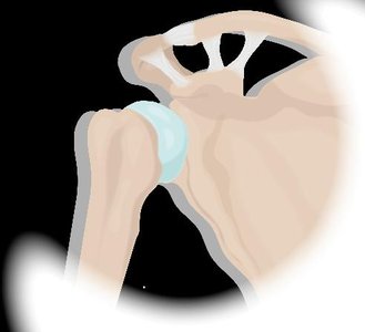
Example: The shoulder joint has a large range of motion but is relatively unstable, making it prone to dislocation. In contrast, the sutures of the skull are immovable and highly stable.
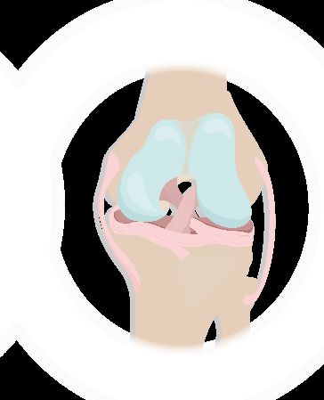
Map of the Lesson on Joints
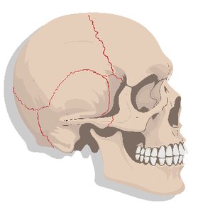
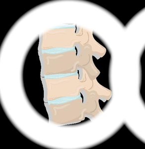
This lesson map provides a visual guide to the classification and structure of joints, helping students navigate the complexity of joint types and their functions.
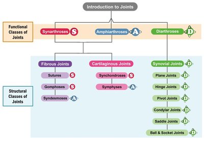
Classification of Joints
Functional Joint Classifications
Joints are classified functionally based on the amount of movement they allow:
Synarthrosis: Immovable or stationary joints (e.g., sutures of the skull).
Amphiarthrosis: Slightly movable joints (e.g., intervertebral joints).
Diarthrosis: Freely movable joints, predominant in the appendicular skeleton (e.g., shoulder, knee).
Rheumatoid arthritis most commonly affects diarthroses, as these joints are dynamic and frequently used.
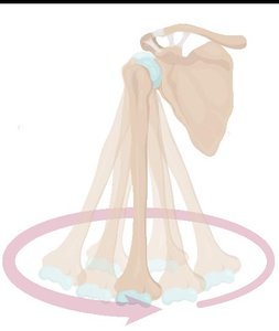
Structural Joint Classifications
Joints are also classified structurally based on the binding material between skeletal elements:
Fibrous Joints: Bound by collagen fibers of dense connective tissue (always synarthrosis or amphiarthrosis).
Cartilaginous Joints: Bound by cartilage (always synarthrosis or amphiarthrosis).
Synovial Joints: Have a synovial cavity and ligaments within an articular capsule (always diarthrosis).
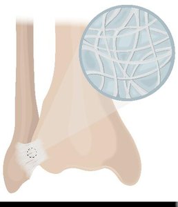
Structural Class: Fibrous Joints
Sutures
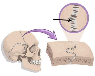
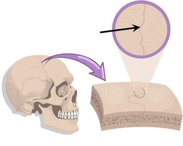
Sutures are fibrous joints of dense irregular connective tissue found only joining bones of the skull. They provide structural protection to the brain but allow no movement. Sutures allow the skull to expand as the brain grows during youth, but ossify in adults, forming a synostosis (bony joint).
Immovable: Sutures are synarthroses.
Ossification: Sutures ossify to form a single bone in adulthood.
Example: Sutures allow for skull expansion in youth and protect the brain.
Gomphosis
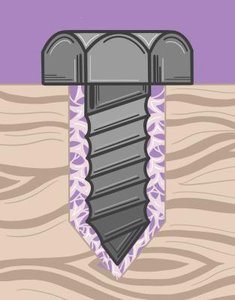
Gomphoses are fibrous joints found only "bolting" teeth to their bony sockets in the gums. The periodontal ligament, made of dense irregular connective tissue, anchors teeth to the jawbone. Gomphoses are immovable except during chewing, and the ligament deteriorates in youth to allow baby teeth to fall out.
Immovable: Gomphoses are synarthroses.
Periodontal Ligament: Anchors teeth to bone.
Example: Gomphosis in the gums.
Syndesmosis
Syndesmoses are fibrous joints connecting bones via ligaments made of dense irregular connective tissue. Greater bone separation and longer fibers allow more mobility than sutures or gomphoses. Interosseous membranes are sheets of dense connective tissue between certain bones (e.g., radius and ulna, tibia and fibula).
Slightly Movable: Syndesmoses are amphiarthroses.
Interosseous Membranes: Provide flexibility and strength.
Example: Syndesmosis between tibia and fibula.
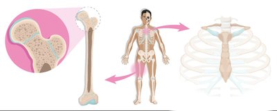
Structural Class: Cartilaginous Joints
Synchondrosis
Synchondroses are cartilaginous joints where bones are bound by hyaline cartilage. They provide structural support but allow no movement. Some synchondroses ossify over time to become synostoses (bony joints).
Immovable: Synchondroses are synarthroses.
Ossification: Hyaline cartilage can be replaced by bone.
Example: Epiphyseal plates and costosternal synchondrosis.
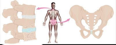
Symphysis
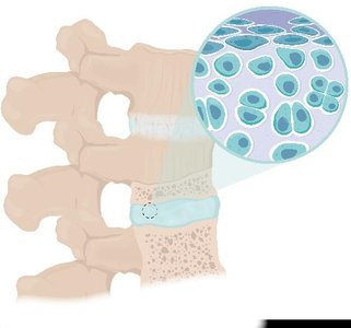
Symphyses are cartilaginous joints in the body's midline where bones are bound by fibrocartilage. They are designed for strength and flexibility, allowing slight movement. Fibrocartilage acts as a shock absorber.
Slightly Movable: Symphyses are amphiarthroses.
Shock Absorption: Fibrocartilage is compressible and resilient.
Example: Intervertebral joints and pubic symphysis.
Structural Class: Synovial Joints
Synovial Joints
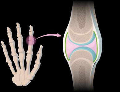
Most joints in the body are synovial joints, which allow free movement and have unique characteristics:
Synovial Cavity: Space between bones storing synovial fluid.
Synovial Fluid: Lubricates and absorbs shock.
Articular Cartilage: Hyaline cartilage covering bone ends, reducing friction.
Articular Capsule: Surrounds the joint with an inner synovial membrane (produces fluid) and an outer fibrous layer (dense connective tissue).
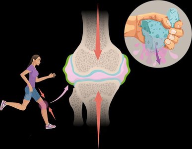
Weeping Lubrication of Synovial Joints
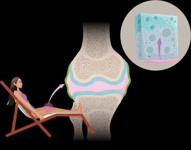
Weeping lubrication is the process where synovial fluid in articular cartilages is forced out upon compression. When pressure is relieved, synovial fluid soaks back in, similar to water in a sponge. This mechanism reduces friction and protects the joint.
Compression: Squeezes synovial fluid out of cartilage.
Decompression: Fluid is reabsorbed into cartilage.
Example: Knee joint during movement.
Additional Features in Some Synovial Joints
Some synovial joints have additional features that enhance their function:
Bursae: Synovial fluid-filled sacs that reduce friction between ligaments/tendons and other tissues.
Tendon Sheath: Elongated bursa wrapping around a tendon, reducing friction.
Fatty Pads: Present in knee and hip joints, providing cushioning.
Articular Discs (Menisci): Discs of fibrocartilage or fat that divide the synovial cavity and act as shock absorbers.