 뒤로
Liver, Gallbladder, and Pancreas: Structure and Function in Digestion
스터디 가이드 - 스마트 노트
 자료에 맞춘 맞춤형 노트, 핵심 정의, 예시, 맥락을 확장해 제공합니다.
Liver, Gallbladder, and Pancreas
Overview of Digestive Functions
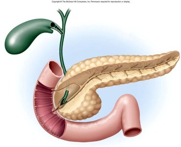
The liver, gallbladder, and pancreas are essential organs that release secretions into the small intestine to facilitate digestion. Their coordinated actions ensure the breakdown and absorption of nutrients.
Liver: Produces bile, metabolizes nutrients, detoxifies blood.
Gallbladder: Stores and concentrates bile.
Pancreas: Secretes digestive enzymes and hormones.
Functions of the Liver
Metabolic and Regulatory Roles
The liver is a multifunctional organ involved in metabolism, detoxification, and storage. It plays a central role in maintaining homeostasis.
Carbohydrate metabolism: Regulates blood glucose by storing glycogen and releasing glucose.
Fat metabolism: Synthesizes cholesterol and lipoproteins; breaks down fatty acids.
Protein metabolism: Deaminates amino acids, produces urea, synthesizes plasma proteins.
Removal of drugs and hormones: Detoxifies blood, metabolizes hormones.
Excretion and synthesis of bile: Bile aids in fat digestion; bile salts are synthesized for emulsification.
Storage: Stores vitamins (A, D, E, K, B12), minerals (iron, copper), and glycogen.
Phagocytosis: Kupffer cells remove old RBCs and WBCs.
Removal of toxins: Detoxifies alcohol, ammonia, and other substances.
Gross Anatomy of the Liver
Lobes and Ligaments
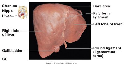
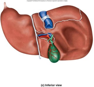
The liver is a large, reddish-brown organ located inferior to the diaphragm. It is divided into four lobes and is supported by several ligaments.
Lobes: Right, left, caudate, and quadrate.
Falciform ligament: Separates right and left lobes.
Round ligament: Remnant of the umbilical vein.
Liver Cirrhosis and Hepatitis
Pathological Changes
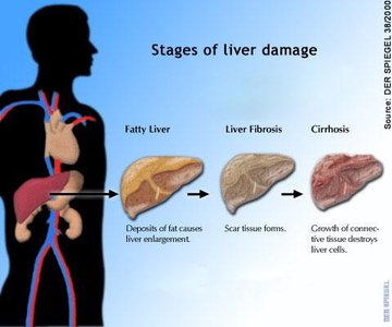
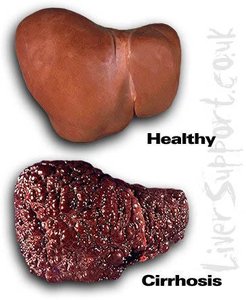
Liver diseases such as cirrhosis and hepatitis affect liver function and structure. Cirrhosis is characterized by scarring and loss of functional tissue, while hepatitis is inflammation caused by viral infection.
Cirrhosis: Progressive scarring (fibrosis) following cell death; impairs liver function.
Stages: Fatty liver → fibrosis → cirrhosis.
Hepatitis: Types A (acute, foodborne), B (blood/body fluid, vaccine available), C (chronic, blood/body fluid, risk of cancer).
Hepatic Circulation
Arterial and Venous Blood Flow
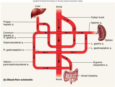
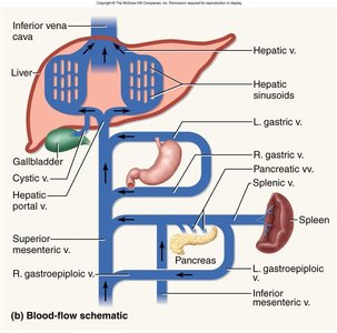
The liver receives blood from both the hepatic artery (oxygen-rich) and the hepatic portal vein (nutrient-rich from the digestive tract). Blood is filtered and then drained via the hepatic veins.
Proper hepatic artery: Supplies oxygenated blood.
Hepatic portal vein: Brings nutrient-rich blood from intestines.
Hepatic vein: Drains filtered blood to the inferior vena cava.
Microscopic Anatomy of the Liver
Hepatic Lobules and Portal Triad
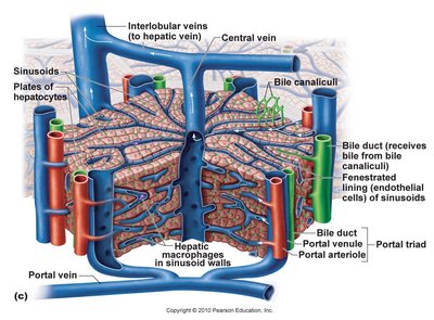
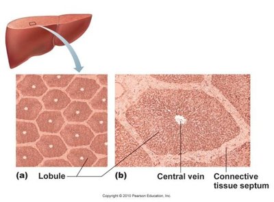
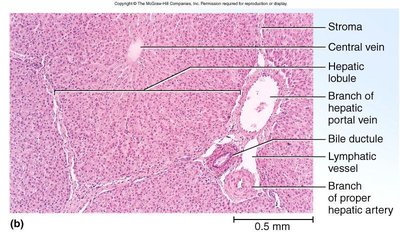
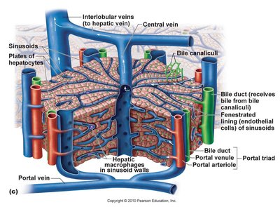
The liver is organized into hexagonal lobules, each containing a central vein surrounded by plates of hepatocytes and sinusoids. The portal triad is found at each corner of the lobule.
Hepatic lobules: Functional units; central vein at the center.
Sinusoids: Capillary spaces where blood is filtered by hepatocytes.
Kupffer cells: Macrophages in sinusoids, remove debris and old cells.
Portal triad: Branch of hepatic portal vein, hepatic artery, and bile duct.
Bile Production and Flow
Biliary Duct System
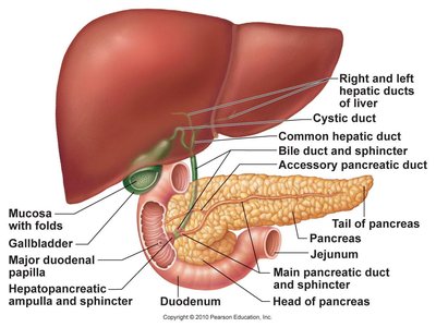
Bile produced by hepatocytes is collected in canaliculi, then flows through bile ducts to the gallbladder or directly to the duodenum. The flow is regulated by the hepatopancreatic sphincter.
Bile canaliculi: Small channels between hepatocytes.
Bile ducts: Collect bile and transport it to the gallbladder or duodenum.
Hepatopancreatic sphincter: Controls release of bile and pancreatic juice.
Gallbladder and Bile
Storage and Function
The gallbladder stores and concentrates bile, which is essential for fat digestion. Bile contains pigments, salts, cholesterol, and phospholipids.
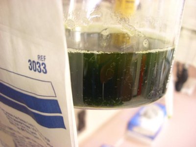
Bile: Yellow-green fluid; emulsifies fats.
Bilirubin: Pigment from hemoglobin breakdown; converted to stercobilin (brown color of feces).
Gallbladder disease: Can cause gray-white, fatty streaked stool.
Gallstones
Formation and Clinical Significance
Gallstones (biliary calculi) form when cholesterol crystallizes due to imbalance between cholesterol and bile salts. They can block bile flow and cause pain.
Risk factors: Female, forty, fertile, fat (high cholesterol diet).
Treatment: Laparoscopic surgery or lithotripsy.
Gross Anatomy of the Pancreas
Structure and Dual Function
The pancreas is a retroperitoneal gland with both endocrine and exocrine functions. It secretes hormones into the blood and digestive enzymes into the duodenum.
Parts: Head, body, tail.
Endocrine: Islets of Langerhans secrete insulin and glucagon.
Exocrine: Acinar cells secrete pancreatic juice (enzymes and bicarbonate).
Pancreatic duct: Opens at hepatopancreatic sphincter.
Hormonal Control of Secretion
Regulation by Cholecystokinin and Secretin
Digestive secretions are regulated by hormones released from the duodenum in response to food.
Cholecystokinin (CCK): Stimulates gallbladder contraction, pancreatic enzyme secretion, and relaxation of sphincter.
Secretin: Stimulates secretion of pancreatic juice, bicarbonate, and bile.
Small Intestine
Structure and Function
The small intestine is the primary site for chemical digestion and nutrient absorption. It consists of three segments: duodenum, jejunum, and ileum.
Duodenum: Neutralizes stomach acid, emulsifies fats, inactivates pepsin, receives pancreatic enzymes.
Jejunum: Most digestion and absorption occur here.
Ileum: Contains Peyer's patches (lymphatic nodules), absorbs bile salts, ends at large intestine.
Surface Area Adaptations
Folds, Villi, and Microvilli
The small intestine has structural adaptations to maximize surface area for absorption.
Circular folds: Slow movement of chyme, increase absorption.
Villi: Fingerlike projections with blood vessels and lacteals.
Microvilli: Brush border enzymes for final digestion.
Brunner's glands: Secrete bicarbonate-rich mucus.
Peyer's patches: Immune function in ileum.
Intestinal Motility
Mixing and Propulsion
Motility in the small intestine involves mixing chyme with digestive juices and moving residue toward the large intestine.
Peristalsis: Wave-like contractions push chyme forward.
Segmentation: Ring-like constrictions mix and churn contents.
Anatomy of the Large Intestine
Segments and Features
The large intestine absorbs water and electrolytes, forms feces, and houses bacterial flora. It consists of several segments and has unique structural features.
Segments: Cecum, appendix, ascending, transverse, descending, sigmoid colon, rectum, anal canal.
Haustra: Pouches formed by teniae coli (longitudinal muscle bands).
Ileocecal valve: Controls entry from ileum.
Microscopic Anatomy of the Large Intestine
Cell Types and Mucosa
The mucosa is primarily simple columnar epithelium, except in the anal canal where it is stratified squamous. There are no villi or circular folds, and mucus is produced by intestinal crypts.
Teniae coli: Three bands of longitudinal muscle.
Haustra: Sacs formed by muscle contraction.
Bacterial Flora
Role in Digestion and Health
The large intestine is populated by diverse bacterial species that ferment undigested carbohydrates and synthesize vitamins.
Fermentation: Produces gases and absorbable sugars.
Vitamin synthesis: B and K vitamins.
Flatus: Gas produced by bacteria and swallowed air.
Absorption and Motility in the Large Intestine
Water Balance and Transit Time
Transit time is 12-24 hours. Water and electrolytes are reabsorbed, and feces are formed. Motility is regulated by haustral contractions.
Diarrhea: Too little water absorbed; rapid transit.
Constipation: Too much water absorbed; slow transit.
Diverticulitis and Colon Cancer
Clinical Conditions
Diverticulitis is inflammation of pouches in the colon wall, often due to low fiber diets. Colon cancer risk increases with polyps, low fiber, high fat, and genetic factors.
Diverticulitis: More common in descending colon.
Colon cancer: Early detection via colonoscopy is key.
Lactose Intolerance
Enzyme Deficiency
Lactose intolerance results from decreased lactase enzyme activity, leading to inability to digest lactose after childhood.
Prevalence: Varies by ethnicity.
Management: Avoid lactose or use enzyme supplements.
Breakdown Products of Digestion
Absorption of Nutrients
Macromolecules are broken down into absorbable units:
Carbohydrates: Monosaccharides
Fats: Glycerol and fatty acids
Proteins: Amino acids
DNA/RNA: Nucleotides
Vitamins: Absorbed unchanged
Carbohydrate Digestion
Enzymatic Breakdown
Starch digestion begins in the mouth and is completed in the small intestine.
Salivary amylase: Inactivated in stomach (pH < 4.5).
Pancreatic amylase: Completes starch digestion.
Brush border enzymes: Final breakdown to monosaccharides.
Protein Digestion
Enzymatic Breakdown
Protein digestion begins in the stomach and continues in the small intestine.
Pepsin: Active in stomach (pH 1.5-3.5), inactivated in duodenum.
Trypsin and chymotrypsin: Digest proteins in small intestine.
Fat Digestion
Absorption Pathways
Fats are emulsified by bile and digested by pancreatic lipases. Most fats are absorbed via lacteals and transported to the liver.
Bile: Emulsifies fats for enzyme action.
Lacteals: Lymphatic vessels absorb fats.
Nucleic Acid Digestion
Enzymatic Breakdown
Nucleic acids are digested by pancreatic nucleases into nucleotides, which are absorbed in the small intestine.
Pancreatic nucleases: Digest DNA and RNA.
