 뒤로
Lymphatic System and Immunity: Anatomy & Physiology Study Guide
스터디 가이드 - 스마트 노트
 자료에 맞춘 맞춤형 노트, 핵심 정의, 예시, 맥락을 확장해 제공합니다.
Lymphatic System and Immunity
Introduction to the Lymphatic System and Immunity
The lymphatic system is a crucial component of the body's defense mechanisms, working alongside the immune system to resist infection and disease. All body cells and tissues contribute to immunity, which is the ability to resist infection and disease.
Immunity: The ability to resist infection and disease.
Immune System: Composed of all cells and tissues involved in immunity.
Functions of the Lymphatic System
The lymphatic system performs several essential functions to maintain homeostasis and protect the body.
Production, maintenance, and distribution of lymphocytes and other lymphoid cells.
Return of excess fluid to the bloodstream.
Maintenance of normal blood volume.
Transport of hormones, nutrients, and wastes.
Components of the Lymphatic System
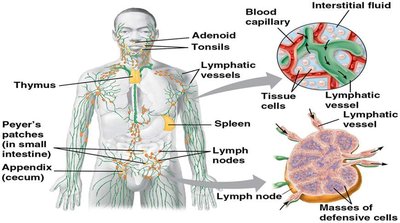
The lymphatic system consists of four main components:
Lymph: A fluid similar to plasma but lacking plasma proteins.
Lymphatic vessels: Carry lymph from peripheral tissues to veins.
Lymphoid tissues and organs: Scattered throughout the body.
Lymphoid cells: Includes lymphocytes, phagocytes, and other cells.
Lymphatic Vessels and Capillaries
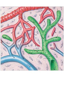
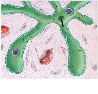
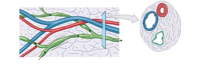
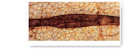
Lymphatic vessels transport lymph from tissues back to the venous system. Lymphatic capillaries are specialized structures that allow the entry of fluids, solutes, viruses, and bacteria, but prevent their return to intercellular spaces.
Lymphatic capillaries: Endothelial cells loosely bound, forming one-way valves.
Structure: Closed at one end, larger luminal diameter, thinner walls than blood vessels.
Lymph flow: From capillaries to larger vessels with one-way valves.
Major Lymphatic Ducts
Lymphatic vessels join to form large trunks that empty into two major collecting vessels:
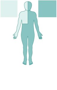
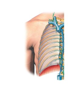
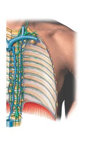
Thoracic duct: Drains lymph from tissues inferior to the diaphragm and the left side of the upper body; empties into the left subclavian vein.
Right lymphatic duct: Drains lymph from the right side of the upper body; empties into the right subclavian vein.
Lymphoid Tissues
Lymphoid tissues are connective tissues dominated by lymphocytes. Lymphoid nodules are areolar tissue packed with lymphocytes, often found in mucosa-associated lymphoid tissue (MALT) in the digestive system.
MALT: Lymphoid tissues associated with the digestive system.
Appendix: Contains a mass of fused lymphoid nodules.
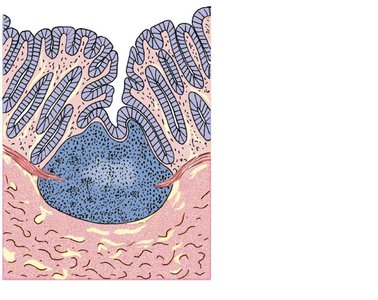
Lymphoid Organs
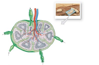
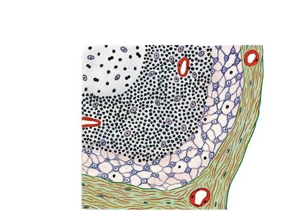
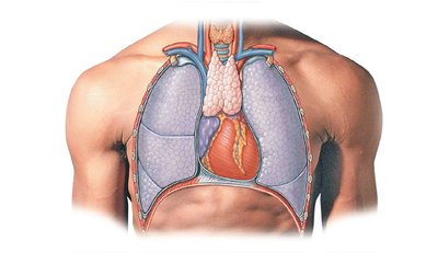
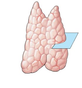
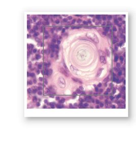
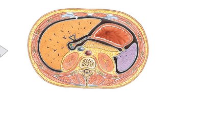
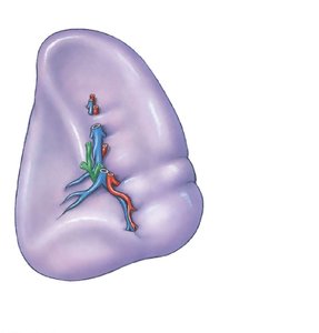
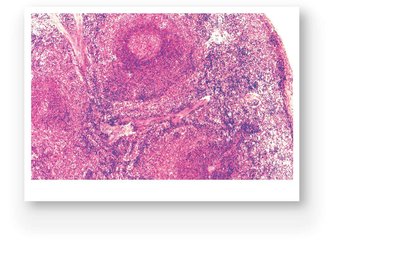
Lymphoid organs are separated from surrounding tissues by a fibrous capsule and include lymph nodes, thymus, and spleen.
Lymph nodes: Purify lymph before it returns to venous circulation, removing 99% of antigens.
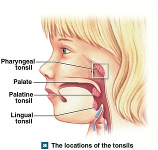
Tonsils: Five tonsils in the pharynx wall; inflammation is called tonsillitis.
Largest lymph nodes: Located in groin, axillae, and base of neck; swell in response to infection.
Lymphocytes
Lymphocytes are a type of leukocyte essential for immune responses. They are produced in bone marrow, thymus, and peripheral lymphoid tissues.
T cells: Thymus-dependent, involved in cell-mediated immunity.
B cells: Bone marrow-derived, responsible for antibody-mediated immunity.
NK cells: Natural killer cells, provide immune surveillance.
Types of Immunity
Immunity is classified as innate (nonspecific) or adaptive (specific).
Innate immunity: Present at birth, works against any type of invading agent.
Adaptive immunity: Acquired after birth, protects against specific pathogens.
Innate Defenses
Innate defenses include physical barriers, phagocytes, immune surveillance, interferons, complement, inflammation, and fever.
Physical barriers: Skin, hair, epithelial layers, secretions.
Phagocytes: Microphages and macrophages that engulf pathogens.
Immune surveillance: NK cells destroy abnormal cells.
Interferons: Chemical messengers that coordinate defenses against viral infections.
Complement: Proteins that assist antibodies in pathogen destruction.
Inflammation: Localized response to injury.
Fever: Elevation of body temperature to accelerate defenses.
Innate Defenses: Immune Surveillance
NK cells carry out immune surveillance by identifying and lysing abnormal cells using perforins.
Steps: Recognition and adhesion, realignment of Golgi apparatus, secretion of perforin, lysis of abnormal cell.
Innate Defenses: Interferons
Interferons are cytokines that trigger production of antiviral proteins and block viral replication.
Interferon alpha (α): Produced by virus-infected cells, attracts and stimulates NK cells.
Interferon beta (β): Secreted by fibroblasts, slows inflammation.
Interferon gamma (γ): Secreted by T cells and NK cells, stimulates macrophage activity.
Innate Defenses: Complement System
The complement system consists of over 30 proteins that assist antibodies in destroying pathogens. Activation occurs via classical, lectin, or alternative pathways.
Classical pathway: Activated by antibodies binding to bacterial cell walls.
Lectin pathway: Activated by mannose-binding lectin binding to carbohydrates on bacterial surfaces.
Alternative pathway: Activated by properdin and other proteins interacting in plasma.
Effects: Cell lysis, enhanced phagocytosis, inflammation.
Innate Defenses: Inflammation and Fever
Inflammation is a localized response to injury, characterized by redness, swelling, heat, and pain. Fever is an elevation of body temperature above 37.2ºC, increasing metabolic rate and inhibiting pathogens.
Products of inflammation: Necrosis, pus, abscess.
Effects: Temporary repair, prevention of pathogen spread, mobilization of defenses.
Adaptive Defenses
Adaptive immunity involves lymphocytes with receptors for specific antigens. When activated, they divide to produce clones. Adaptive defenses are coordinated by T cells and B cells.
Cell-mediated immunity: Provided by cytotoxic T cells, defends against abnormal cells and pathogens inside cells.
Antibody-mediated immunity: Provided by B cells, defends against antigens and pathogens in body fluids.
Properties of Adaptive Immunity
Specificity: Each T or B cell responds only to a specific antigen.
Versatility: Many types of lymphocytes, each fighting different antigens.
Memory: Inactive lymphocytes (memory cells) provide immunity against new exposure.
Tolerance: Immune system ignores normal antigens (self-antigens).
Adaptive Defenses: T Cells
T cells are subdivided into cytotoxic, helper, regulatory, and memory T cells. Activation requires antigen recognition and costimulation.
Cytotoxic T cells: Attack antigens physically and chemically.
Helper T cells: Stimulate responses of T cells and B cells.
Regulatory T cells: Moderate immune response.
Memory T cells: Respond to previously encountered antigens.
Adaptive Defenses: B Cells
B cells differentiate into plasma cells that produce antibodies. Sensitization and activation require antigen binding and helper T cell costimulation.
Plasma cells: Synthesize and secrete antibodies.
Memory B cells: Remain in reserve for future infections.
Antibody Structure and Function
Antibodies are soluble proteins composed of two pairs of polypeptide chains (heavy and light), each with constant and variable segments. The variable segments determine antigen specificity.
Antigen-binding site: Region that binds to antigenic determinant sites (epitopes).
Antigen–antibody complex: Complete antigens have at least two determinant sites; exposure leads to B cell sensitization and immune response.
Actions of antibodies: Neutralization, precipitation, agglutination, complement activation, phagocyte attraction, inflammation stimulation, prevention of adhesion.
Hapten: Partial antigens that must attach to carrier molecules to act as complete antigens.
Primary and Secondary Immune Responses
Primary response occurs after first exposure to antigen, taking time to develop. Secondary response is faster and more extensive due to memory cells.
Immunocompetence and Immune Disorders
Immunocompetence is the ability to produce an immune response after antigen exposure. Hypersensitivities (allergies) are excessive responses to antigens. Autoimmune disorders occur when the system fails to recognize self-antigens, leading to attacks on normal cells.
Examples: Thyroiditis, rheumatoid arthritis, type 1 diabetes.
Effects of Aging and System Integration
The immune system diminishes with age, increasing vulnerability to infections and cancer. The nervous and endocrine systems influence immune responses.
Summary Table: Types of Lymphocytes
Type | Origin | Main Function |
|---|---|---|
T cells | Thymus | Cell-mediated immunity |
B cells | Bone marrow | Antibody-mediated immunity |
NK cells | Bone marrow | Immune surveillance |
Summary Table: Types of Immunity
Type | Characteristics | Example |
|---|---|---|
Innate | Present at birth, nonspecific | Skin barrier |
Adaptive | Acquired, specific | Antibody production |
Summary Table: Actions of Antibodies
Action | Description |
|---|---|
Neutralization | Blocks antigen binding sites |
Precipitation/Agglutination | Forms immune complexes |
Complement Activation | Destroys pathogens |
Phagocyte Attraction | Enhances phagocytosis |
Inflammation Stimulation | Promotes inflammatory response |
Prevention of Adhesion | Blocks pathogen attachment |
Additional info: Academic context and expanded explanations were added to ensure completeness and clarity for exam preparation.
