 뒤로
뒤로Lymphatic System and Immunity: Structured Study Notes
스터디 가이드 - 스마트 노트
 자료에 맞춘 맞춤형 노트, 핵심 정의, 예시, 맥락을 확장해 제공합니다.
Lymphatic System and Immunity
Overview of the Lymphatic System
The lymphatic system is a network of vessels, tissues, and organs that play a crucial role in fluid balance, lipid absorption, and immune defense. It is closely associated with the cardiovascular system and is essential for maintaining homeostasis and protecting the body from pathogens.
Transport of Excess Interstitial Fluid: Removes excess fluid from tissues and returns it to the bloodstream.
Absorption of Lipids: Specialized lymphatic capillaries called lacteals absorb dietary lipids from the digestive tract.
Defense Against Disease: Provides immune surveillance and permanent immunity against infections.
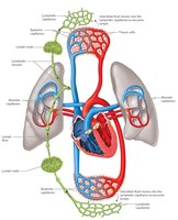
Lymphatic Pathways
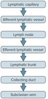
Lymphatic pathways consist of a series of vessels and nodes that transport lymph from tissues back to the circulatory system. The flow follows a hierarchical structure:
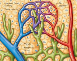
Lymphatic Capillaries: Microscopic, closed-ended tubes that collect interstitial fluid, now called lymph.
Lymphatic Vessels: Thin-walled vessels with semilunar valves, similar to veins, that transport lymph toward lymph nodes.
Lymph Nodes: Filter lymph and house immune cells.
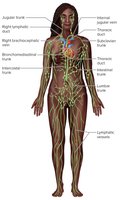
Lymphatic Trunks: Drain lymph from regions of the body.
Collecting Ducts: Two main ducts (thoracic and right lymphatic duct) return lymph to the subclavian veins.
Lymphatic Trunks and Collecting Ducts
Thoracic Duct: Drains most of the body; begins as the cisterna chyli and empties into the left subclavian vein.
Right Lymphatic Duct: Drains the upper right portion of the body; empties into the right subclavian vein.
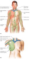
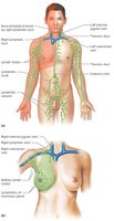
Tissue Fluid and Lymph
Lymph is formed when tissue fluid enters lymphatic capillaries. Tissue fluid is derived from blood plasma, minus large plasma proteins, which remain in the blood to maintain osmotic pressure.
Formation: Filtration from plasma exceeds reabsorption, creating tissue fluid. Increased hydrostatic pressure forces fluid into lymphatic capillaries.
Prevention of Edema: Lymphatic drainage prevents accumulation of excess tissue fluid.
Lymph Flow
Low Hydrostatic Pressure: Lymph flow is aided by skeletal muscle contraction, respiratory movements, smooth muscle contraction, and valves.
Highest During Exercise: Physical activity increases lymph flow.
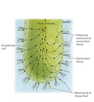
Lymphatic Tissues and Organs
Lymphatic tissues contain lymphocytes and macrophages. Organs include lymph nodes, thymus, and spleen.
Lymph Nodes
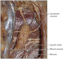
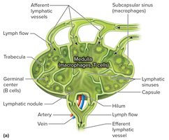
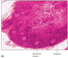
Structure: Bean-shaped, <2.5 cm long, located along lymphatic vessels.
Function: Filter pathogens, house lymphocytes and macrophages.
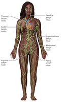
Locations: Cervical, axillary, supratrochlear, inguinal, pelvic, abdominal, thoracic regions.
Thymus
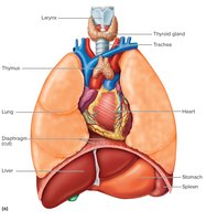
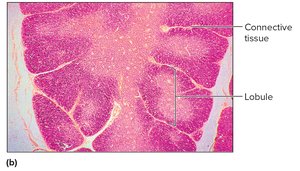
Location: Mediastinum, divided into lobules.
Function: Maturation of T lymphocytes; produces thymosins.
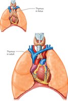
Size Changes: Large in infancy, shrinks at puberty, replaced by adipose tissue in elderly.
Spleen
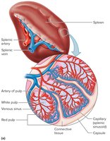
Largest Lymphatic Organ: Located in upper left abdomen.
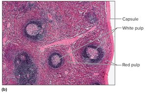
Structure: Contains white pulp (lymphocytes, macrophages) and red pulp (RBCs, macrophages).
Function: Filters blood, breaks down worn-out RBCs, immune defense.
Immunity: Body Defenses Against Infection
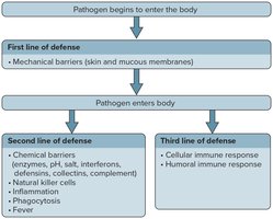
The lymphatic system defends against pathogens through innate and adaptive mechanisms.
Innate (Nonspecific) Defenses: General defenses against many pathogens.
Adaptive (Specific) Defenses: Target specific antigens; carried out by lymphocytes.
Innate Defenses
Mechanical Barriers: Skin, mucous membranes.
Inflammation: Redness, swelling, heat, pain; walls off infection.
Chemical Barriers: Enzymes, interferons, defensins, collectins, complement system.
Natural Killer Cells: Lyse virus-infected and cancer cells.
Phagocytosis: Neutrophils and monocytes engulf pathogens.
Fever: Elevated temperature inhibits microbial growth.
Adaptive Defenses
Antigens: Non-self molecules that evoke immune response.
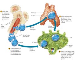
Lymphocyte Origins: T cells mature in thymus; B cells mature in bone marrow.
T Cells and Cellular Immune Response
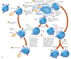
Activation: Requires antigen-presenting cells (APCs) displaying antigen fragments bound to MHC proteins.
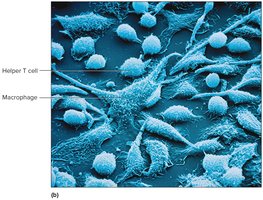
Types: Helper T cells, cytotoxic T cells, memory T cells, regulatory T cells.
B Cells and Humoral Immune Response
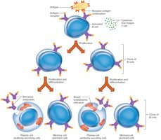
Activation: Antigen binds to B cell receptor; cytokines from T cells stimulate proliferation.
Plasma Cells: Secrete antibodies (immunoglobulins).
Memory B Cells: Provide future immunity.
Antibody Molecules
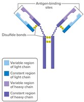
Structure: Y-shaped, composed of 2 heavy and 2 light chains; variable and constant regions.
Types: IgG, IgA, IgM, IgD, IgE.
Antibody Actions
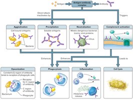
Direct Attack: Agglutination, precipitation, neutralization.
Complement Activation: Opsonization, chemotaxis, lysis, inflammation.
Immune Responses
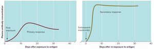
Primary Response: First exposure to antigen; slower, lower antibody concentration.
Secondary Response: Subsequent exposure; rapid, high antibody concentration.
Hypersensitivity Reactions
Type I (Immediate): Allergy; IgE-mediated, histamine release.
Type II: Antibody-dependent cytotoxic reaction.
Type III: Immune-complex reaction.
Type IV: Delayed-reaction hypersensitivity.
Transplantation and Autoimmunity
Transplant Types: Autografts, isografts, allografts, xenografts.
Tissue Rejection: Immune response against foreign antigens; immunosuppressive drugs used.
Autoimmunity: Immune system attacks self tissues; production of autoantibodies.
Life-Span Changes
Immune Decline: Thymus shrinks, immune response weakens with age.
Antibody Changes: IgA and IgG increase; IgM and IgE decrease.
Autoantibodies: Increased risk of autoimmune diseases in elderly.
