 뒤로
Membrane Transport & Excitable Cells: Synaptic Transmission and Muscle Contraction
스터디 가이드 - 스마트 노트
 자료에 맞춘 맞춤형 노트, 핵심 정의, 예시, 맥락을 확장해 제공합니다.
Membrane Transport & Excitable Cells
The Synapse: Where Electricity Becomes Chemistry
Synapses are specialized junctions where neurons communicate with other neurons, muscle fibers, or glands. This process is fundamental to nervous system function and muscle contraction.
Presynaptic neuron: Sends the signal.
Postsynaptic cell: Receives the signal; can be another neuron, muscle fiber, or gland.
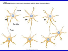
Types of synaptic arrangements:
Axodendritic: Most common, often on dendritic spines.
Axosomatic: Onto the cell body.
Axoaxonic: Modulates transmitter release from the target terminal.
Electrical vs Chemical Synapses
Neurons communicate via two main types of synapses: electrical and chemical.
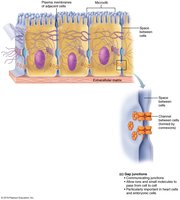
Electrical synapses:
Gap junctions directly connect cytoplasm of adjacent cells.
Very fast, can be bidirectional.
Synchronize activity (e.g., heart, smooth muscle).
Chemical synapses:
Cells separated by a synaptic cleft (30–50 nm).
Neurotransmitter released from presynaptic neuron binds to receptors on postsynaptic cell.
Slower, but highly modifiable.
Events at a Chemical Synapse
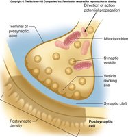
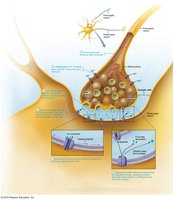
Chemical synaptic transmission involves several steps:
Action potential arrives at axon terminal.
Voltage-gated Ca2+ channels open; Ca2+ enters.
Ca2+ triggers exocytosis of synaptic vesicles.
Neurotransmitter diffuses across cleft and binds receptors.
Ion channels open, causing a graded postsynaptic potential.
Signal ends via reuptake, enzymatic breakdown, or diffusion.
Synaptic Delay
Chemical synapses introduce a delay (0.3–5 ms), mainly due to Ca2+ entry and vesicle fusion. This delay is the rate-limiting step in neural processing.
Postsynaptic Potentials: EPSPs and IPSPs
Postsynaptic potentials are graded changes in membrane potential:
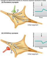
Excitatory postsynaptic potential (EPSP): Depolarizes membrane, moving toward threshold.
Inhibitory postsynaptic potential (IPSP): Hyperpolarizes membrane, moving away from threshold.
Mechanisms of EPSP and IPSP
Glutamate: Main Excitatory Neurotransmitter
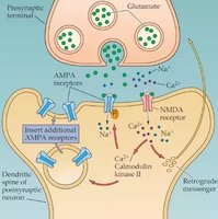
Glutamate binds ligand-gated cation channels, allowing Na+ influx and K+ efflux. The net effect is depolarization (EPSP).
GABA and Glycine: Main Inhibitory Neurotransmitters
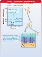
GABA (brain) and glycine (spinal cord) open K+ or Cl- channels, causing hyperpolarization (IPSP).
Synaptic Integration: Summation
Neurons integrate multiple inputs through summation:
Temporal summation: One input fires rapidly; EPSPs stack in time.
Spatial summation: Multiple inputs fire together; EPSPs add across space.
EPSPs and IPSPs add algebraically; inhibition can cancel excitation.
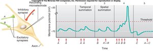
Axon Initial Segment: The Decision Point
The axon initial segment has the lowest threshold due to the highest density of voltage-gated Na+ channels. Graded potentials decay with distance, so synapses closer to the initial segment have more influence.
Neurotransmitters: The Chemical Vocabulary of the Nervous System
Classification by Chemical Structure
Neurotransmitters are grouped into six families:
Acetylcholine (ACh): Neuromuscular junctions, autonomic nervous system, attention, memory.
Biogenic amines: Dopamine, norepinephrine, epinephrine, serotonin, histamine.
Amino acids: Glutamate (excitatory), GABA & glycine (inhibitory).
Peptides: Substance P (pain), endorphins, enkephalins (natural opiates).
Purines: ATP, adenosine.
Gases & lipids: Nitric oxide, CO, H2S, endocannabinoids.
Classification by Function
Effect: Excitatory or inhibitory depends on the receptor, not the molecule.
Action:
Direct (ionotropic): Receptor is an ion channel; fast, brief.
Indirect (metabotropic): G protein–coupled receptor; slower, broader, longer-lasting.
Drugs, Toxins, and the Chemical Synapse
Agonist: Binds and activates the receptor.
Antagonist: Binds and blocks the receptor.
Neural Circuits: Patterns of Synaptic Connection
Four Types of Neural Circuits
Diverging: One neuron amplifies and spreads the signal to many.
Converging: Many neurons concentrate the signal to one.
Reverberating: Feedback loop for rhythmic, sustained output.
Parallel after-discharge: Parallel paths reconverge for a burst of output.
Nerve Meets Muscle: The Neuromuscular Junction (NMJ)
Anatomy of the NMJ
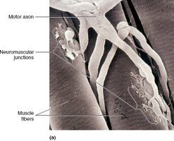
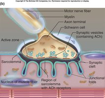
The NMJ is the synapse between a motor neuron and a skeletal muscle fiber. Each muscle fiber has one NMJ, located about midway along its length.
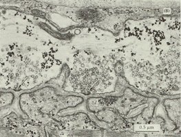
Axon terminals: Contain synaptic vesicles with acetylcholine (ACh).
Synaptic cleft: Filled with basal lamina.
Motor end plate: Highly folded sarcolemma rich in nicotinic ACh receptors (nAChRs).
Role of Nicotinic ACh Receptors
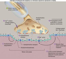
nAChRs are ligand-gated cation channels. When ACh binds, Na+ enters and K+ exits, creating an end plate potential (EPP) that triggers a muscle action potential.
Events at the NMJ: Six Steps to a Muscle Action Potential
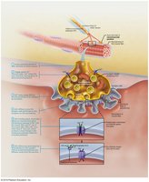
AP arrives at axon terminal of motor neuron.
Voltage-gated Ca2+ channels open; Ca2+ enters.
Ca2+ causes ACh release by exocytosis.
ACh diffuses across cleft and binds ACh receptors on sarcolemma.
Ligand-gated channels open; Na+ and K+ pass, creating EPP.
ACh broken down by acetylcholinesterase and diffuses away.
Clinical Connections: NMJ Disorders
Botulinum toxin: Blocks ACh release, causing flaccid paralysis.
Curare: Blocks nicotinic receptors, causing paralysis.
Myasthenia gravis: Antibodies destroy ACh receptors, causing muscle weakness.
Nerve agents (e.g., sarin): Inhibit acetylcholinesterase, preventing muscle relaxation.



Grading the Force: Motor Units, Twitches, and Summation
The Motor Unit
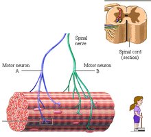
A motor unit consists of one motor neuron and all the muscle fibers it innervates. All fibers in a unit contract together, and units are scattered throughout the muscle.
The Muscle Twitch
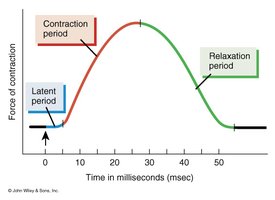
A muscle twitch is a single contraction in response to a single stimulus, consisting of three phases:
Latent period: E-C coupling; no tension.
Contraction: Cross-bridge cycling; tension peaks.
Relaxation: Ca2+ reuptake; tension falls.
Temporal Summation: Frequency
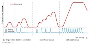
Increasing stimulus frequency leads to summation:
Single twitch: Full relaxation between stimuli.
Wave summation: Stimulus before relaxation; tension adds.
Unfused tetanus: High frequency; quivering contraction.
Fused tetanus: Very high frequency; smooth, maximal contraction.
Spatial Summation: Recruitment
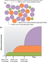
Recruitment activates more motor units, increasing force. Small units are recruited first for fine control; large units are added for power.
Isotonic vs Isometric Contractions
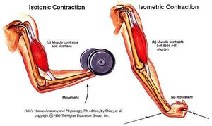
Isotonic: Muscle changes length (concentric: shortens; eccentric: lengthens).
Isometric: Muscle length stays the same; tension builds but does not exceed the load.

Muscle Fibre Types, Metabolism, and Fatigue
Three Ways to Make ATP
Creatine phosphate: CP + ADP → creatine + ATP; lasts ~15 s.
Anaerobic glycolysis: Glucose → pyruvic acid → lactic acid; 2 ATP/glucose; lasts ~30–40 s.
Aerobic respiration: Mitochondria; ~32 ATP/glucose; requires O2; lasts hours.


Muscle Fibre Types
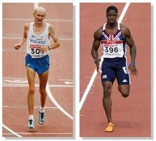
Slow oxidative: Endurance, fatigue-resistant.
Fast oxidative-glycolytic: Intermediate speed and fatigue resistance.
Fast glycolytic: Fast, powerful, fatigues quickly.
Fibre Types in Real Life
Most muscles are a mix of fiber types, with proportions largely determined by genetics. Training can shift fiber properties.
Smooth Muscle: Structure and Function
Location and Layers
Smooth muscle is found in the walls of hollow organs (digestive, urinary, reproductive tracts, airways, blood vessels). Usually two layers: longitudinal and circular.
Microscopic Anatomy
No sarcomeres; filaments run diagonally.
Dense bodies anchor thin filaments.
Works over a wide range of lengths.
Innervation and Response
Innervated by autonomic nervous system via varicosities and diffuse junctions.
Response depends on receptor type, hormones, local chemicals, and stretch.
Excitation–Contraction Coupling
Ca2+ enters from ECF and SR.
Ca2+ binds calmodulin (no troponin).
Ca2+-calmodulin activates myosin light chain kinase (MLCK).
MLCK phosphorylates myosin, enabling contraction.
Unitary vs Multiunit Smooth Muscle
Unitary (visceral): Most hollow organs; gap junctions; contract as a unit.
Multiunit: Large airways, arteries, arrector pili, iris; independent fibers, own nerve supply.
Special Features of Smooth Muscle Contraction
Slow and sustained contraction.
Economical energy use; rarely fatigues.
Stress-relaxation: adapts to stretch.
Hyperplasia: cells can divide (e.g., uterus in pregnancy).
Comparison: Skeletal vs Smooth Muscle
Feature | Skeletal Muscle | Smooth Muscle |
|---|---|---|
Striations | Present | Absent |
Control | Voluntary (somatic) | Involuntary (autonomic) |
Contraction speed | Fast | Slow |
Fatigue | Can fatigue | Rarely fatigues |
Cell division | None | Possible (hyperplasia) |
Summary of Key Concepts
Ca2+ triggers neurotransmitter release; receptor type determines effect; signal ends by reuptake, breakdown, or diffusion.
EPSPs and IPSPs sum in time and space; the axon initial segment decides whether to fire.
Six neurotransmitter families; effects depend on receptor and action (direct/indirect).
Four neural circuit types: diverging, converging, reverberating, parallel after-discharge.
Muscle force graded by frequency (tetanus) and recruitment; isotonic vs isometric contractions.
Muscle fiber types match metabolism to function; smooth muscle trades speed for economy.
