 뒤로
Modulation of Contraction and Conduction in Cardiac and Smooth Muscle
스터디 가이드 - 스마트 노트
 자료에 맞춘 맞춤형 노트, 핵심 정의, 예시, 맥락을 확장해 제공합니다.
Muscle Tissue
Modulation of Contraction and Conduction in Cardiac Muscle
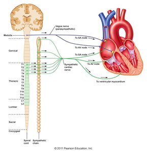
Cardiac muscle contraction and conduction are tightly regulated by both intrinsic properties and neuronal input. The heart's autorhythmic cells generate action potentials, but their activity is modulated by the autonomic nervous system, which influences heart rate and conduction velocity.
Neuronal Input: The heart receives input from both sympathetic and parasympathetic neurons, which modify the rate and force of contraction.
Autorhythmic Cells: These cells, primarily located in the SA node, have an intrinsic firing rate (~90 action potentials per minute) but are modulated to a resting heart rate of 70-72 BPM by parasympathetic dominance.
Tonic Control: Heart rate is under tonic control, meaning it can be adjusted up or down by autonomic input.
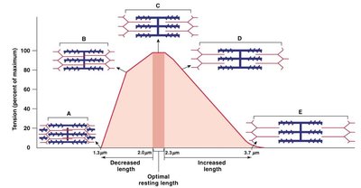
Length-Tension Relationship in Cardiac and Skeletal Muscle
The length-tension relationship describes how muscle tension varies with sarcomere length. In cardiac muscle, stretching the sarcomere increases Ca2+ sensitivity and tension, while in skeletal muscle, optimal overlap between thick and thin filaments maximizes force production.
Stretched Sarcomere: Increases Ca2+ sensitivity and entry, enhancing contraction.
Overlap: Degree of overlap between filaments determines force output.
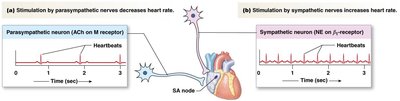
Autonomic Modulation of Heart Rate
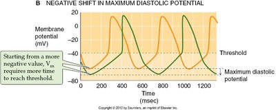
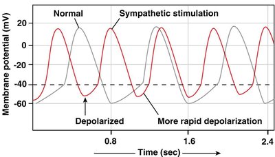
Heart rate is modulated by the balance between sympathetic and parasympathetic activity. Parasympathetic neurons (ACh) decrease heart rate, while sympathetic neurons (NE or epinephrine) increase it.
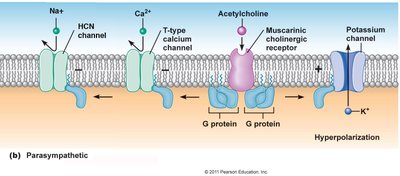
Parasympathetic Effects: ACh acts on muscarinic (M2) receptors, opening K+ channels and closing T-type Ca2+ and HCN channels, leading to hyperpolarization and decreased action potential frequency.
Sympathetic Effects: NE/epinephrine acts on β1 adrenergic receptors, increasing Na+ and Ca2+ conductance, causing faster depolarization and increased heart rate.
Sympathetic Signaling Pathway
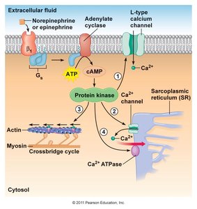
Sympathetic stimulation increases heart rate and contractility via β1 adrenergic receptors, activating a cascade that increases Ca2+ influx and enhances cross-bridge cycling.
Key Steps: NE/epinephrine → β1 receptor → Gs protein → adenylate cyclase → cAMP → protein kinase → increased Ca2+ channel activity.
Smooth Muscle
Structure and Functional Differences
Smooth muscle is structurally and functionally distinct from skeletal and cardiac muscle. It is more variable, operates over a range of lengths, and can sustain contraction without fatigue.
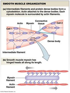
Cell Structure: Spindle-shaped, uninucleate cells; troponin and T-tubules absent; intermediate filaments and dense bodies form cytoskeletal structure.
Contraction: Initiated electrically or chemically; controlled by autonomic nervous system; Ca2+ from extracellular space and/or SR initiates contraction.
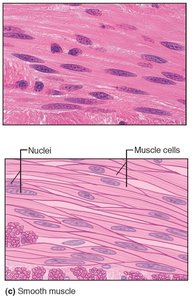
Functional Variability of Smooth Muscle
Classification by Location
Smooth muscle is found in various locations throughout the body, each with specialized functions.
Vascular: Blood vessel walls
Gastrointestinal: Digestive tract and associated organs
Urinary: Bladder and ureters
Respiratory: Airway passages
Reproductive: Uterus and other reproductive structures
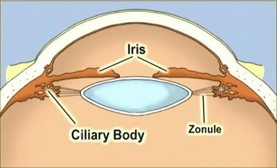
Ocular: Iris and ciliary body
Classification by Contraction Pattern
Smooth muscle contraction patterns can be phasic or tonic, depending on their physiological role.
Phasic: Usually relaxed, contracts periodically (e.g., esophagus).
Phasic Cycling: Cycles between contraction and relaxation (e.g., intestine).
Tonic: Usually contracted, relaxes to allow material passage (e.g., sphincter).
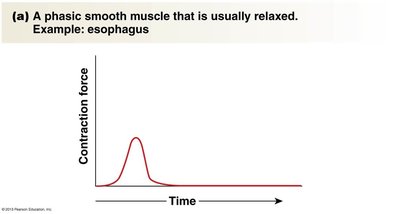
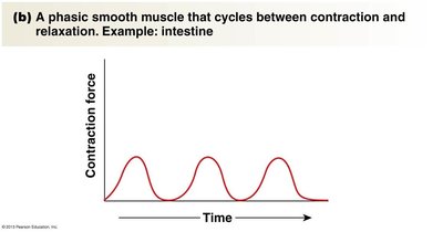
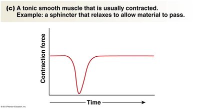
Classification by Communication with Neighboring Cells
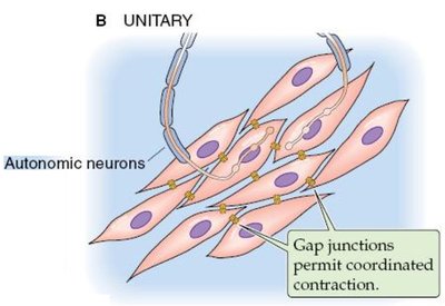
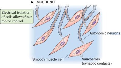
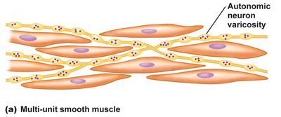
Smooth muscle can be categorized based on how cells communicate and coordinate contraction.
Unitary (Single Unit): Contains gap junctions, allowing coordinated contraction of many cells; found in visceral organs.
Multiunit: Cells are not electrically coupled, allowing finer motor control; found in iris, ciliary body, male reproductive tract, and uterus (except just prior to labor).
Electrical Activity in Smooth Muscle
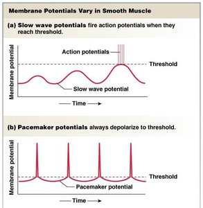
Smooth muscle action potentials are slower than those in skeletal or cardiac muscle, due to Ca2+ channels propagating the AP. Repolarization is also slower, and APs are usually seen in unitary smooth muscle.
Initiation: Can be neural, hormonal, or mechanical.
Spontaneous Activity: Some smooth muscle cells exhibit spontaneous membrane potential oscillations, leading to tonic contractions without action potentials.
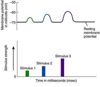
Graded Response: Local depolarizations can trigger Ca2+ entry and contraction.
Ca2+ Initiated Contraction in Smooth Muscle
Contraction in smooth muscle is triggered by increased cytosolic Ca2+, which can occur via several mechanisms:
Voltage-Gated Channels: Ca2+ entry through voltage-gated channels.
SR Release: Ca2+ release from the sarcoplasmic reticulum via RyR (Ca2+-induced Ca2+ release) or IP3R (IP3-mediated release).
Voltage-Independent Channels: Ligand-gated, store-operated, and stretch-activated Ca2+ channels.
Summary Table: Types of Smooth Muscle
Type | Electrical Coupling | Location | Function |
|---|---|---|---|
Unitary (Single Unit) | Gap junctions | Visceral organs (GI, bladder) | Coordinated contraction |
Multiunit | None | Iris, ciliary body, reproductive tract | Fine motor control |
Summary Table: Contraction Patterns in Smooth Muscle
Pattern | State | Example |
|---|---|---|
Phasic | Usually relaxed | Esophagus |
Phasic Cycling | Cycles between contraction and relaxation | Intestine |
Tonic | Usually contracted | Sphincter |
Key Equations
Muscle Tension: $ \text{Tension} = \text{Force generated by cross-bridge cycling} $
Membrane Potential: $ \text{Vm} = \text{Resting membrane potential} + \text{Depolarization} $
Additional info: Academic context was added to clarify the mechanisms of contraction, electrical activity, and classification of smooth muscle types.
