 뒤로
Muscle Anatomy: Structure, Function, and Classification
스터디 가이드 - 스마트 노트
 자료에 맞춘 맞춤형 노트, 핵심 정의, 예시, 맥락을 확장해 제공합니다.
Muscle Anatomy Introduction
Overview of the Muscular System
The muscular system refers specifically to skeletal muscles, which are specialized contractile tissues responsible for movement and stability in the human body. Muscles constitute approximately 40% of body weight, with around 600 skeletal muscles present. The study of skeletal muscles is known as myology. All muscle types convert chemical energy (ATP) into mechanical energy for motion.
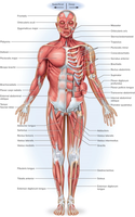
Functions of the Muscular System
Movement: Muscles contract to pull on bones, producing voluntary movement.
Stability: Muscles stabilize joints and maintain posture, including antigravity muscles that resist gravity.
Heat Production (Thermogenesis): Skeletal muscles generate 20-30% of body heat at rest and up to 85% during exercise (e.g., shivering).
Types of Muscle Tissue
Classification and Characteristics
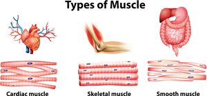
There are three main types of muscle tissue, each with distinct structural and functional properties:
Skeletal Muscle: Voluntary, striated, attached to bones via tendons, controlled by the somatic nervous system.
Cardiac Muscle: Involuntary, striated, found only in the heart, regulated by the autonomic nervous system.
Smooth Muscle: Involuntary, non-striated, located in walls of organs (e.g., intestines, blood vessels).
Structural Organization of Skeletal Muscle
Hierarchy of Muscle Structure
Skeletal muscle is organized from large to small structures:
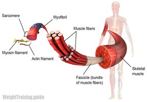
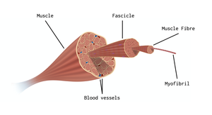
Whole Muscle → Muscle Fascicles → Muscle Fibers (Cells) → Myofibrils → Sarcomeres
Muscle Fiber and Myofibril Structure
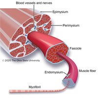
Muscle fibers are long, cylindrical cells containing multiple nuclei. Each fiber houses numerous myofibrils, which are composed of repeating units called sarcomeres—the contractile units responsible for muscle contraction.
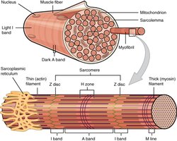
Connective Tissue Components of Skeletal Muscle
Connective Tissue Layers
Skeletal muscle includes several connective tissue layers that provide structure and support:
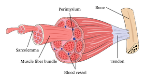
Epimysium: Surrounds the entire muscle.
Perimysium: Surrounds each muscle fascicle (bundle).
Endomysium: Surrounds individual muscle fibers (cells).
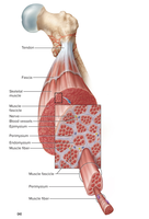
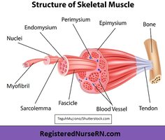
Muscle Attachments
Types of Muscle Attachment
Muscles attach to bones in two primary ways:
Direct Attachment: Epimysium is fused to the bone's periosteum; little separation between muscle and bone.
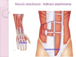
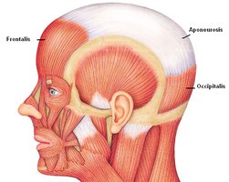
Indirect Attachment: Muscle connective tissue extends to form a tendon (rope-like) or aponeurosis (sheet-like), which attaches to bone.
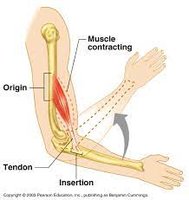
Muscle Points of Attachment: Origin and Insertion
Each muscle attaches to bone at two points:
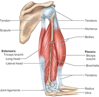
Origin: Stationary, immovable, or less movable attachment (anchor).
Insertion: More movable attachment; moves toward the origin during contraction.
Muscle Actions and Functional Groups
Categories of Muscle Action
Muscles are classified by their actions during movement:
Prime Mover (Agonist): Main muscle responsible for a movement.
Antagonist: Opposes or reverses a movement.
Synergist: Assists the prime mover or reduces unwanted movement.
Fixator: Stabilizes a bone or joint, preventing movement.
Fascicle Arrangement
Types of Fascicle Orientation
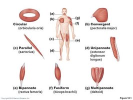
The arrangement of fascicles determines muscle strength and direction of pull:
Circular: Surround external openings (e.g., orbicularis oris).
Convergent: Broad origin, narrow insertion (e.g., pectoralis major).
Parallel: Fascicles run parallel to the long axis (e.g., sartorius).
Fusiform: Thick in the middle, tapered at ends (e.g., biceps brachii).
Pennate: Short fascicles attach to a tendon; includes unipennate, bipennate, and multipennate (e.g., deltoid).
Naming Muscles
Muscle Naming Patterns
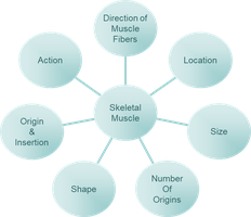
Muscle names often describe their location, shape, size, direction, origins, or actions. Understanding these patterns aids in identifying and visualizing muscles.
Location: Named for nearby structures (e.g., frontalis near frontal bone).
Shape: Describes muscle shape (e.g., deltoid = triangular, trapezius = trapezoid).
Size: Indicates relative size (e.g., maximus = largest, minimus = smallest).
Direction of Fibers: Describes fiber orientation (e.g., rectus abdominis = straight, obliques = angled).
Action: Named for movement performed (e.g., flexor, extensor, adductor).
Combined Categories: Names may combine multiple descriptors (e.g., flexor carpi ulnaris).
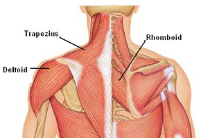
Muscle Pairs and Movement
Muscle Pair Functionality
Muscles are grouped in pairs on the skeleton. Muscles contract and pull bones; they do not push. When one muscle in a pair contracts, its partner relaxes, and vice versa, allowing coordinated movement and return to original position.
Summary Table: Connective Tissue Layers of Skeletal Muscle
Layer | Location | Function |
|---|---|---|
Epimysium | Surrounds entire muscle | Protects and supports muscle |
Perimysium | Surrounds fascicles | Bundles muscle fibers |
Endomysium | Surrounds individual fibers | Provides structural support |
Summary Table: Types of Muscle Tissue
Type | Control | Location | Appearance |
|---|---|---|---|
Skeletal | Voluntary | Attached to bones | Striated |
Cardiac | Involuntary | Heart | Striated |
Smooth | Involuntary | Organs, vessels | Non-striated |
Key Equations
Muscle Contraction Energy Conversion
Muscle contraction involves the conversion of chemical energy (ATP) to mechanical energy:
Additional info:
Muscle contraction is regulated by the nervous system, with skeletal muscle under voluntary control and cardiac/smooth muscle under involuntary control.
Muscle fascicle arrangement affects both the force and range of motion of a muscle.
