 뒤로
Muscle Contraction: Structure, Neuromuscular Junction, and Excitation-Contraction Coupling L13
스터디 가이드 - 스마트 노트
 자료에 맞춘 맞춤형 노트, 핵심 정의, 예시, 맥락을 확장해 제공합니다.
Muscle Tissue Structure
Muscle Fiber Anatomy
Muscle fibers (myocytes) are highly specialized cells responsible for contraction. Their unique structure allows for efficient force generation and transmission.
Sarcolemma: The cell membrane of a muscle fiber, crucial for conducting action potentials.
Sarcoplasm: The cytoplasm of the muscle cell, containing organelles and dissolved ions.
Myofibrils: Cylindrical structures composed of repeating units called sarcomeres, the functional contractile units.
Sarcoplasmic Reticulum (SR): Specialized smooth endoplasmic reticulum that stores and releases calcium ions (Ca2+).
Mitochondria: Provide ATP for contraction.
Nucleus: Muscle fibers are multinucleated, supporting high protein synthesis.
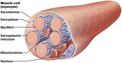
Neuromuscular Junction (NMJ)
Structure and Function
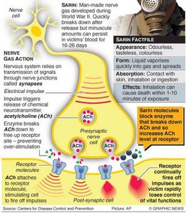
The neuromuscular junction is the synapse between a motor neuron and a muscle fiber, enabling communication for muscle contraction.
Motor Neuron: The presynaptic cell that releases the neurotransmitter acetylcholine (ACh).
Axon Terminal: Contains vesicles filled with ACh.
Synaptic Cleft: The gap between the neuron and muscle fiber.
Motor End Plate: Specialized region of the sarcolemma with ACh receptors.
Acetylcholinesterase (AChE): Enzyme that breaks down ACh, terminating the signal.
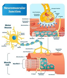
Events at the Neuromuscular Junction
Muscle contraction is initiated by a series of events at the NMJ:
An action potential arrives at the axon terminal, triggering exocytosis of ACh into the synaptic cleft.
ACh binds to receptors on the motor end plate, opening sodium channels and causing depolarization.
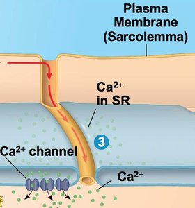
The action potential travels along the sarcolemma and into T-tubules.
Depolarization of T-tubules causes the release of Ca2+ from the terminal cisternae of the SR.
When ACh is degraded by AChE, the signal ends and relaxation begins.
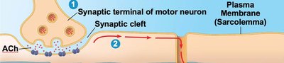
Membrane Potentials & Muscle Contraction
Resting and Action Potentials
Muscle fibers maintain a resting membrane potential, which is essential for excitability and contraction.
Resting Membrane Potential: The inside of the cell is more negative than the outside.
Action Potential: A brief reversal of membrane charge, acting as an electrical signal.
Depolarization: Na+ enters the cell, making the inside more positive.
Repolarization: K+ leaves the cell, restoring the negative potential.
Sodium-Potassium Pump
The Na+/K+ pump maintains the resting membrane potential by moving sodium out and potassium in.
Function: Helps maintain the electrochemical gradient necessary for action potentials.
Equation: per ATP hydrolyzed
Excitation-Contraction Coupling
Calcium Connection
Excitation-contraction coupling links the electrical signal from the neuron to the mechanical contraction of the muscle fiber.
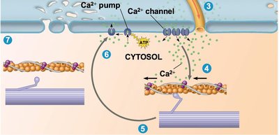
Ca2+ Release: Depolarization of T-tubules triggers Ca2+ release from the SR.
Triad: Consists of two terminal cisternae and one T-tubule.
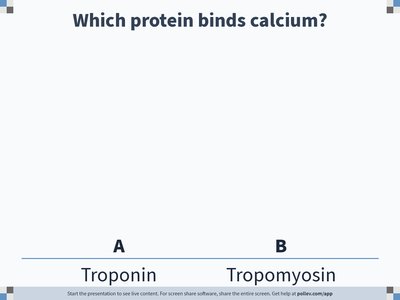
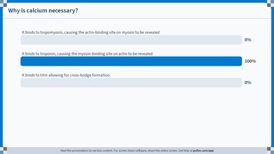
Ca2+ Role: Binds to troponin, initiating contraction.
Regulatory Proteins: Troponin & Tropomyosin
Control of Cross-Bridge Cycling
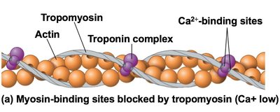
Troponin and tropomyosin regulate when myosin can bind to actin, controlling muscle contraction.
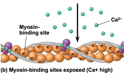
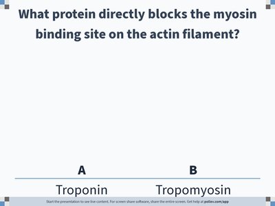
At Rest: Tropomyosin covers actin's myosin-binding sites; troponin holds tropomyosin in place.
During Contraction: Ca2+ binds to troponin, causing a conformational change that shifts tropomyosin and exposes binding sites.
Cross-Bridge Cycling
Steps of the Cycle
Cross-bridge cycling is the process by which myosin heads interact with actin to produce muscle contraction.
Grab: Myosin binds to actin's exposed myosin-binding site, forming a cross-bridge.
Pull (Power Stroke): Myosin head pivots, pulling actin toward the M line and releasing ADP and phosphate.
Release: ATP binds to myosin, causing it to detach from actin.
Reset: Myosin hydrolyzes ATP to ADP and phosphate, re-cocking its head for another cycle.
Requirements for Contraction & Relaxation
Contraction
Muscle contraction requires the presence of acetylcholine, calcium ions, and ATP.
Acetylcholine: Must remain in the synaptic cleft to continue stimulating the motor end plate.
Ca2+: Must be available in the sarcoplasm to bind troponin.
ATP: Required for cross-bridge cycling and myosin reactivation.
Relaxation
Relaxation occurs when acetylcholinesterase degrades acetylcholine, Ca2+ is pumped back into the SR, and ATP detaches cross-bridges.
Acetylcholinesterase: Terminates the signal by breaking down ACh.
Ca2+ Pump: Returns Ca2+ to the SR, ending contraction.
ATP: Detaches myosin from actin and resets the head.
Muscle Tension & Twitch
Generation of Muscle Tension
Muscle tension is the force produced during contraction. It depends on the number of cross-bridges, fiber length, and frequency of stimulation.
Maximum Tension: Achieved when the maximum number of cross-bridges are formed.
Resting Length: Optimal overlap of actin and myosin is necessary (Goldilocks principle).
Frequency of Stimulation: Determines the type of contraction (twitch, treppe, wave summation, tetanus).
Types of Muscle Contractions
Type | Stimulus Timing | Tension | Reason |
|---|---|---|---|
Twitch | Single action potential | Brief contraction | One contraction per AP |
Treppe | Next stimulus after relaxation | Step-wise increase | Ca2+ not fully returned to SR |
Wave Summation | Next stimulus before end of relaxation | Additive, increases | Fibers partially contracted |
Incomplete Tetanus | Next stimulus after start of relaxation | Progressive, plateau | Mostly prevents relaxation |
Complete Tetanus | Next stimulus before relaxation | Maximum, smooth | Prevents relaxation |
Muscle Fatigue
Muscle fatigue occurs when tension declines due to O2 depletion, lactic acid accumulation, and insufficient ATP for Na+/K+ pump.
O2 Depletion: Limits ATP production.
Lactic Acid: Accumulates during anaerobic respiration.
ATP Shortage: Impairs repolarization and contraction.
Summary Table: Key Terms and Functions
Term | Definition | Function |
|---|---|---|
Sarcolemma | Muscle cell membrane | Conducts action potentials |
Sarcoplasmic Reticulum | Specialized ER in muscle | Stores/releases Ca2+ |
Troponin | Regulatory protein | Binds Ca2+, shifts tropomyosin |
Tropomyosin | Regulatory protein | Blocks myosin-binding sites on actin |
Acetylcholine (ACh) | Neurotransmitter | Initiates muscle contraction |
Acetylcholinesterase (AChE) | Enzyme | Breaks down ACh |
ATP | Energy molecule | Drives cross-bridge cycling |
Example: During a sprint, rapid action potentials at the NMJ cause repeated Ca2+ release, leading to sustained contraction and eventual muscle fatigue due to O2 depletion.
