 뒤로
Muscle Tissue and Physiology: Structure, Function, and Mechanisms
스터디 가이드 - 스마트 노트
 자료에 맞춘 맞춤형 노트, 핵심 정의, 예시, 맥락을 확장해 제공합니다.
Muscle Tissue Overview
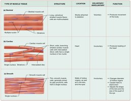
Types of Muscle Tissue
Muscle tissue is specialized for contraction and force generation, and is essential for movement, posture, and regulation of internal processes. There are three main types of muscle tissue: skeletal, cardiac, and smooth muscle.
Skeletal muscle: Long, multinucleated, striated fibers; voluntary control; attached to skeleton for movement.
Cardiac muscle: Short, branched, striated cells; involuntary control; found only in the heart.
Smooth muscle: Spindle-shaped, non-striated cells; involuntary control; lines hollow organs and vessels.
Functions of Muscle Tissue
Movement: Muscles contract to move body parts or propel substances.
Posture: Maintains body position.
Joint stabilization: Supports and stabilizes joints.
Heat generation: Muscle activity produces heat.
Regulation of flow: Smooth muscle controls passage through hollow organs.
Properties and Structure of Muscle Cells
Properties of Muscle Cells
Contractility: Ability to contract and generate tension.
Excitability: Responds to stimuli.
Conductivity: Conducts electrical signals across membrane.
Extensibility: Can be stretched without damage.
Elasticity: Returns to original shape after stretching.
Structure of Muscle Cells
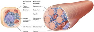
Muscle cells (fibers) are specialized compared to generalized cells, containing unique organelles for contraction.
Sarcolemma: Plasma membrane of muscle cell.
Sarcoplasm: Cytoplasm of muscle cell.
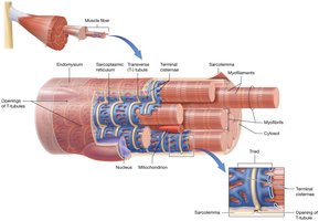
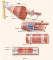
Myofibrils: Bundles of contractile proteins.
Sarcoplasmic reticulum (SR): Stores and releases calcium ions.
Transverse tubules (T-tubules): Extensions of sarcolemma; conduct action potentials.
Structure and Function of Skeletal Muscle Fibers
Skeletal Muscle Fiber Anatomy
Skeletal muscle fibers are long, cylindrical cells formed by fusion of myoblasts, resulting in multiple nuclei. They are surrounded by endomysium and can be up to 30 cm in length.

Myofibrils and Myofilaments
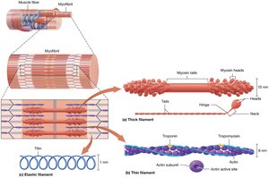
Myofibrils are composed of myofilaments, which are classified as:
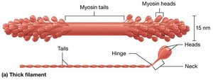
Thick filaments: Made of myosin; heads bind actin and hydrolyze ATP.
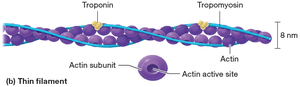
Thin filaments: Made of actin, tropomyosin, and troponin; actin provides binding sites for myosin.
Elastic filaments: Made of titin; provide structural support and elasticity.
Sarcomere Structure
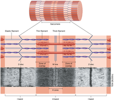
The sarcomere is the functional unit of muscle contraction, defined by specific bands and lines:
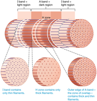
I band: Contains only thin filaments.
Z disc: Anchors thin and elastic filaments; marks sarcomere boundaries.
A band: Contains both thick and thin filaments; zone of overlap.
H zone: Contains only thick filaments.
M line: Anchors thick filaments.
Levels of Organization in Skeletal Muscle
Muscle fibers are grouped into fascicles, which are bundled to form muscles. Connective tissue layers (endomysium, perimysium, epimysium) provide support and connect muscles to bones via tendons.
Muscle Disease: Duchenne Muscular Dystrophy (DMD)
DMD Pathology
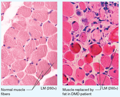
DMD is a degenerative disease caused by a defective gene for dystrophin, leading to breakdown of muscle fibers and replacement with fat and connective tissue.
Symptoms: Weakness, waddling gait, early wheelchair use, early death from respiratory/cardiac failure.
Sliding-Filament Mechanism of Contraction
Mechanism of Contraction
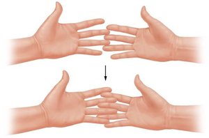
Muscle contraction occurs as myosin heads pull actin filaments toward the M line, shortening the sarcomere without changing filament length.
I band and H zone: Narrow during contraction.
A band: Remains unchanged.
Myosin heads: Attach, pull, release, and reset in a cycle.
Membrane Potentials and Muscle Excitability
Resting Membrane Potential (RMP)
Muscle fibers maintain a resting membrane potential due to unequal ion distribution, primarily Na+ and K+, across the sarcolemma.
RMP: Typically -85 mV in skeletal muscle.
Na+/K+ pump: Maintains gradients by moving 3 Na+ out and 2 K+ in per ATP hydrolyzed.
Ion Channels
Leak channels: Always open; allow passive ion movement.
Gated channels: Open in response to stimuli (ligand, voltage, mechanical).
Electrochemical Gradients
K+: Concentration gradient favors exit; electrical gradient favors entry.
Na+: Both gradients favor entry.
Action Potentials
Action potentials are rapid changes in membrane potential, essential for muscle contraction.
Depolarization: Na+ enters, membrane becomes less negative.
Repolarization: K+ exits, membrane returns to resting state.
Skeletal Muscle Contraction and Relaxation
Neuromuscular Junction (NMJ)
The NMJ is the synapse between a motor neuron and muscle fiber, where acetylcholine (ACh) triggers muscle excitation.
Axon terminal: Releases ACh.
Synaptic cleft: Space between neuron and muscle.
Motor end plate: Contains ligand-gated Na+ channels.
Phases of Contraction
Excitation phase: ACh binds to channels, Na+ enters, end-plate potential generated.
Excitation-contraction coupling: Action potential travels along sarcolemma and T-tubules, Ca2+ released from SR.
Contraction phase: Ca2+ binds troponin, tropomyosin moves, myosin binds actin, crossbridge cycle begins.
Muscle Relaxation
Acetylcholinesterase: Degrades ACh, channels close.
Ca2+ pumped back into SR: Troponin and tropomyosin block actin, muscle relaxes.
Energy Sources for Muscle Contraction
ATP Requirements
Na+/K+ pumps
Myosin head detachment and recocking
Ca2+ reuptake into SR
ATP Generation
Immediate: Stored ATP and creatine phosphate (10 seconds).
Glycolytic: Glycolysis (30–40 seconds); anaerobic, produces lactic acid.
Oxidative: Aerobic catabolism in mitochondria; sustained ATP production.
Muscle Tension and Fiber Types
Twitch Contraction
Latent period: Action potential propagation.
Contraction period: Crossbridge cycling.
Relaxation period: Ca2+ removal.
Wave Summation and Tetanus
Unfused tetanus: Partial relaxation between stimuli.
Fused tetanus: No relaxation; maximal tension.
Length-Tension Relationship
Optimal sarcomere length (100–120% of natural length) allows maximal tension generation.
Fiber Types
Type I (slow-twitch): Small diameter, oxidative, fatigue-resistant, dark muscle.
Type II (fast-twitch): Large diameter, glycolytic, fatigues quickly, white muscle.
Type IIa: Intermediate, oxidative-glycolytic.
Type IIx: Fast glycolytic, powerful, fatigues quickly.
Fiber Type | Diameter | ATPase Activity | Energy Source | Fatigue Resistance |
|---|---|---|---|---|
Type I | Small | Low | Oxidative | High |
Type IIa | Intermediate | High | Oxidative/Glycolytic | Moderate |
Type IIx | Large | High | Glycolytic | Low |
Muscle Tension at the Organ Level
Motor Units and Muscle Tone
Motor unit: One motor neuron and all fibers it innervates.
Recruitment: Activation of more motor units increases tension.
Muscle tone: Baseline involuntary activation for posture and readiness.
Types of Muscle Contractions
Isotonic concentric: Muscle shortens, tension constant.
Isotonic eccentric: Muscle lengthens, tension constant.
Isometric: Muscle length unchanged, tension generated.
Muscle Performance and Adaptation
Myoplasticity
Endurance training: Increases mitochondria, blood supply, fatigue resistance.
Resistance training: Increases fiber diameter and myofibrils (hypertrophy).
Disuse: Decreases fiber size and strength (atrophy).
Muscular Fatigue
Depletion of ATP sources
Decreased oxygen
Accumulation of metabolic byproducts
Environmental stress
Excess Postexercise Oxygen Consumption (EPOC)
Restores homeostasis after exercise
Replenishes oxygen, restores ion balance, dissipates heat
Smooth and Cardiac Muscle
Smooth Muscle Structure and Function
Propels materials, forms sphincters, regulates flow
No striations; actin and myosin arranged obliquely
Dense bodies anchor filaments
Contraction via Ca2+-calmodulin-MLCK pathway
Single-unit (coordinated) and multi-unit (independent) types
Cardiac Muscle Structure and Function
Short, branched, striated cells
Intercalated discs for electrical and physical connection
Autorhythmic pacemaker cells coordinate contraction
Challenge Questions
What anchors thin and elastic filaments in place within the myofibril? Answer: B) Z disc
What is accomplished by the power stroke? Answer: D) The power stroke results in myosin heads pulling actin toward the center of the sarcomere.
What does the depolarization of the transverse tubules (T-tubules) promote? Answer: B) Release of calcium from the sarcoplasmic reticulum
Key Equations
Na+/K+ Pump: Maintains ion gradients across sarcolemma
Resting Membrane Potential:
Summary Table: Muscle Fiber Types
Type | Diameter | ATPase Activity | Energy Source | Fatigue Resistance |
|---|---|---|---|---|
Type I | Small | Low | Oxidative | High |
Type IIa | Intermediate | High | Oxidative/Glycolytic | Moderate |
Type IIx | Large | High | Glycolytic | Low |
Additional info: This guide expands on brief points with academic context, definitions, and examples for clarity and completeness.
