 뒤로
Muscle Tissue and Physiology: Structured Study Notes
스터디 가이드 - 스마트 노트
 자료에 맞춘 맞춤형 노트, 핵심 정의, 예시, 맥락을 확장해 제공합니다.
Muscle Tissue and Physiology
Types of Muscle Tissue
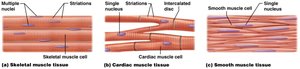
Muscle tissue is classified into three main types, each with distinct structural and functional characteristics. All muscle types generate force, known as muscle tension, which is essential for movement and stability.
Skeletal muscle: Voluntary, striated, multinucleated cells; responsible for body movement.
Cardiac muscle: Involuntary, striated, single nucleus per cell, intercalated discs; found only in the heart.
Smooth muscle: Involuntary, non-striated, single nucleus; lines hollow organs and blood vessels.
Functions of Muscle Tissue
Muscle tissue performs several vital functions in the body:
Movement: Facilitates movement of the skeleton and internal organs.
Maintain posture: Keeps the body upright and stable.
Stabilize joints: Supports and reinforces joints.
Regulate flow: Controls passage of materials through hollow organs (e.g., blood vessels, intestines).
Properties of Muscle Cells
Muscle cells (myocytes) possess unique properties that enable their function:
Contractility: Ability to contract and generate force.
Excitability: Ability to respond to stimuli.
Conductivity: Ability to conduct electrical signals across the membrane.
Extensibility: Ability to stretch without damage.
Elasticity: Ability to return to original shape after stretching.
Structure of Muscle Cells
Muscle Cell Anatomy
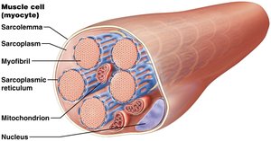
Muscle cells contain specialized structures for contraction and energy production:
Sarcoplasm: Cytoplasm of the muscle cell.
Sarcolemma: Plasma membrane of the muscle cell.
Sarcoplasmic reticulum (SR): Modified endoplasmic reticulum that stores calcium ions.
Myofibrils: Cylindrical organelles responsible for contraction.
Mitochondria: Provide energy for contraction.
Structure of the Skeletal Muscle Fiber
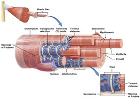
Skeletal muscle fibers have specialized features for efficient contraction:
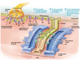
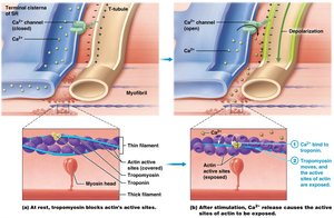
Transverse tubules (T-tubules): Inward extensions of the sarcolemma that surround myofibrils.
Terminal cisternae: Enlarged sections of SR flanking each T-tubule.
Triad: Consists of two terminal cisternae and one T-tubule.
Structure of the Myofibril and Myofilaments
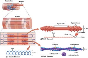
Myofibrils are composed of myofilaments, which are categorized by their function:
Thick filaments: Made of myosin (contractile protein).
Thin filaments: Made of actin (contractile), tropomyosin and troponin (regulatory proteins).
Elastic filaments: Made of titin (structural protein).
The Sarcomere and Muscle Contraction
Sarcomere Structure
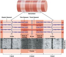
The sarcomere is the functional unit of muscle contraction, defined by specific bands and lines:
I band: Contains only thin filaments.
Z disc: Structural proteins in the middle of the I band.
A band: Contains both thick and thin filaments.
H zone: Middle of the A band, only thick filaments.
M line: Structural proteins holding thick filaments in place.
Sliding Filament Theory
The sliding filament theory explains how muscle contraction occurs:
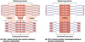
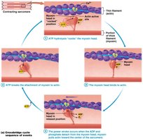
Myosin heads attach to actin and pull thin filaments toward the M line.
Z-discs move closer together, shortening the sarcomere.
Filaments themselves do not shorten; thin filaments slide past thick filaments.
Muscle Cell Electrophysiology
Membrane Potential
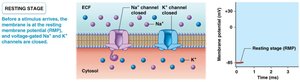
Muscle cells maintain a resting membrane potential due to unequal ion distribution:
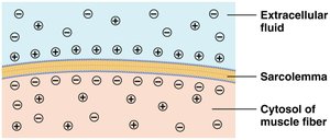
Resting membrane potential is typically -85 mV.
Layer of anions in the intracellular fluid (ICF) and cations in the extracellular fluid (ECF) creates an electrical gradient.
Na+/K+ Pump
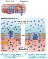
The Na+/K+ pump maintains the concentration gradients of sodium and potassium:
Moves 3 Na+ ions out and 2 K+ ions into the cell.
Requires ATP hydrolysis.
High Na+ in ECF, high K+ in cytosol.
Action Potentials
Action potentials are rapid changes in membrane potential that trigger muscle contraction:
Generated by opening gated ion channels.
Ligand-gated channels open in response to chemicals; voltage-gated channels open in response to changes in membrane potential.
Depolarization: Na+ channels open, Na+ enters, membrane potential becomes less negative.
Repolarization: K+ channels open, K+ exits, membrane potential returns to resting value.
Neuromuscular Junction and Muscle Contraction
Neuromuscular Junction (NMJ)
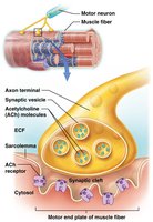
The NMJ is the site where a motor neuron communicates with a muscle fiber:
Motor unit: Motor neuron and all connected muscle fibers.
Synapse: Junction between neuron and muscle cell.
Synaptic cleft: Space between axon terminal and muscle fiber.
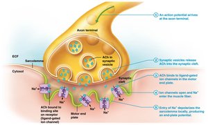
Acetylcholine (ACh): Neurotransmitter released from synaptic vesicles.
Motor end plate: Region of sarcolemma with ligand-gated Na+ channels.
Phases of Skeletal Muscle Contraction
Muscle contraction occurs in three phases:
Excitation phase: Action potential arrives at NMJ, ACh released, Na+ channels open.
Excitation-contraction coupling: Action potential propagates along sarcolemma and T-tubules, Ca2+ released from SR.
Contraction phase: Crossbridge cycle begins, myosin heads pull actin filaments.
Skeletal Muscle Relaxation
Relaxation occurs when:
ACh is degraded by acetylcholinesterase.
Ligand-gated Na+ channels close, sarcolemma returns to resting potential.
Ca2+ is pumped back into SR, troponin and tropomyosin block actin sites.
Rigor Mortis
Rigor mortis is the postmortem stiffening of muscles due to lack of ATP, preventing detachment of myosin from actin.
Muscle Energy and Twitch Contraction
Energy Requirements
ATP is essential for:
Na+/K+ pump function
Myosin head detachment and recocking
Pumping Ca2+ back into SR
Sources of Energy
Immediate: Stored ATP and creatine phosphate (10 seconds)
Glycolytic catabolism: Glucose/glycogen (30-40 seconds)
Oxidative catabolism: Mitochondria (longer duration)
Twitch Contraction
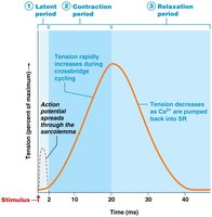
A muscle twitch is the smallest unit of contraction, consisting of three phases:
Latent period: AP propagates across sarcolemma.
Contraction period: Crossbridge cycling generates tension.
Relaxation period: Ca2+ pumped back into SR, tension decreases.
Length-Tension Relationship
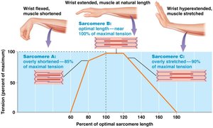
Optimal sarcomere length (100–120% of natural length) allows maximal tension generation due to maximal crossbridge formation.
Types of Muscle Fibers and Motor Units
Classes of Skeletal Muscle Fibers
Type I (slow-twitch): Small diameter, slow contraction, high fatigue resistance, oxidative metabolism, high myoglobin, red color.
Type II (fast-twitch): Large diameter, fast contraction, low fatigue resistance, glycolytic metabolism, low myoglobin, white color.
Motor Units
Slow motor units: Type I fibers, fine control.
Fast motor units: Type II fibers, powerful contractions.
Small motor units: Few fibers, precise movements (e.g., fingers).
Large motor units: Many fibers, less control, more power (e.g., legs).
Recruitment: Activation of additional motor units for increased force.
Muscle tone: Baseline involuntary activation for posture and readiness.
Types of Muscle Contractions
Isotonic and Isometric Contractions
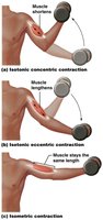
Isotonic concentric: Muscle shortens.
Isotonic eccentric: Muscle lengthens.
Isometric: Muscle length remains unchanged.
Muscle Adaptation and Fatigue
Changes Caused by Physical Training
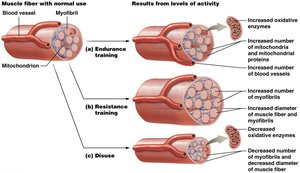
Endurance training: Increases blood supply, mitochondria, oxidative enzymes, and fatigue resistance.
Resistance training: Increases myofibrils and fiber diameter (hypertrophy).
Disuse: Decreases myofibrils, fiber size, and oxidative enzymes (atrophy).
Muscular Fatigue and Recovery
Fatigue: Inability to maintain contraction intensity; caused by metabolite depletion, oxygen shortage, chemical accumulation, and environmental factors.
Excess Postexercise Oxygen Consumption (EPOC): Increased breathing rate post-exercise to restore homeostasis.
Smooth and Cardiac Muscle
Smooth Muscle
Functions: Peristalsis, sphincter formation, regulation of flow.
Structure: No sarcomeres, calmodulin replaces troponin, myosin heads along entire thick filament, less extensive SR, no T-tubules.
Types: Single unit (gap junctions, most common), multi-unit (independent contraction, less common).
Cardiac Muscle
Structure: Striated, single nucleus, intercalated discs (gap junctions and desmosomes).
Function: Contracts as a coordinated unit, contains pacemaker cells, autorhythmic (spontaneously generates action potentials).
