 뒤로
Muscle Tissue I: Structure, Function, and Physiology
스터디 가이드 - 스마트 노트
 자료에 맞춘 맞춤형 노트, 핵심 정의, 예시, 맥락을 확장해 제공합니다.
Muscle Tissue: Types and Characteristics
Major Functions and Structural Elements
Muscle tissue is specialized for contraction and is essential for movement, stability, and various physiological functions. There are three main types of muscle tissue: skeletal, cardiac, and smooth muscle, each with unique structural and functional characteristics.
Skeletal muscle: Long, multinucleated, striated fibers; voluntary control; attached to bones; responsible for body movement.
Cardiac muscle: Short, branched, uni- or binucleated cells; striated; involuntary; found in the heart; connected by gap junctions; responsible for pumping blood.
Smooth muscle: Short, spindle-shaped, uninucleated cells; non-striated; involuntary; found in walls of hollow organs; controls diameter and movement of substances.
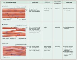
Key Characteristics of Muscle Cells
Contractility: Ability of protein fibers within myocytes to draw together and produce tension.
Excitability: Responds to electrical or chemical stimuli.
Conductivity: Conducts electrical stimulus along the cell membrane.
Extensibility: Can be stretched up to three times its resting length.
Elasticity: Regains original shape after stretching.
Organization of Skeletal Muscle
Arrangement of Skeletal Myocytes and Tissues
Skeletal muscle is organized into hierarchical structures that allow efficient contraction and force transmission.
Endomysium: Surrounds individual muscle cells (myocytes).
Perimysium: Groups muscle cells into fascicles.
Epimysium: Encloses the entire muscle; continues as tendon.
Tendon: Connects muscle to bone, transmitting force.
Structure of Skeletal Muscle Fibers
Cell Size, Shape, and Internal Components
Skeletal muscle fibers are large, cylindrical, multinucleated cells with visible striations. They contain specialized structures for contraction and energy production.
Sarcoplasm: Cytoplasm of the muscle cell.
Sarcolemma: Plasma membrane of the muscle cell.
Sarcoplasmic reticulum (SR): Modified endoplasmic reticulum; stores and releases calcium ions.
Myofibrils: Bundles of contractile proteins; responsible for striations and contraction.
Mitochondria: Provide energy for contraction.
Nuclei: Multiple per cell, located at the periphery.

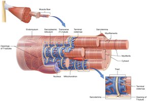
Transverse Tubules and Terminal Cisternae
Transverse tubules (T-tubules): Deep extensions of the sarcolemma; surround each myofibril and allow rapid transmission of action potentials.
Terminal cisternae: Enlarged sections of the SR; flank each T-tubule; together with T-tubule form a triad.
Myofibrils and Myofilaments
Types of Myofilaments
Thick filaments: Composed of myosin.
Thin filaments: Composed of actin, tropomyosin, and troponin.
Elastic filaments: Composed of titin; provide structural support and elasticity.
Arrangement in Sarcomeres
Myofilaments are organized into repeating units called sarcomeres, which are the functional contractile units of muscle fibers.
I band: Contains only thin filaments; appears light.
Z disc: Anchors thin and elastic filaments; marks the boundary of a sarcomere.
A band: Contains both thick and thin filaments; appears dark.
H zone: Central region of A band with only thick filaments.
M line: Middle of A band; anchors thick filaments.
Sliding Filament Mechanism of Contraction
During contraction, actin filaments slide along myosin filaments toward the M line, pulling Z-discs closer together and shortening the sarcomere.
Relaxed sarcomere: Wide I band and H zone.
Contracted sarcomere: Narrowed I band and H zone; A band remains unchanged.
Mechanism: Myosin heads bind to actin, pull, and release in a cycle powered by ATP.
Membrane Potential in Skeletal Myocytes
Definition and Generation of Resting Membrane Potential
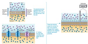
The membrane potential is the electrical potential difference across the sarcolemma, resulting from the separation of charged particles. The resting membrane potential in skeletal myocytes is typically -85 mV.
Electrical gradient: Separation of ions across the membrane.
Voltage: Difference in electrical potential between two points.
Resting membrane potential: Maintained by ion channels and pumps, especially the sodium-potassium pump.
Sodium-Potassium Pump
Function: Actively transports 3 Na+ out and 2 K+ into the cell, using ATP.
Result: High [Na+] outside, high [K+] inside; creates and maintains the negative resting membrane potential.
Relevant Equation
The resting membrane potential can be estimated using the Nernst equation:
Where E is the equilibrium potential, R is the gas constant, T is temperature, z is the charge of the ion, and F is Faraday's constant.
Summary Table: Muscle Tissue Types
Type | Structure | Location | Voluntary/Involuntary | Function |
|---|---|---|---|---|
Skeletal | Long, multinucleated, striated fibers | Attached to skeleton | Voluntary | Movement of the body |
Cardiac | Short, branched, striated, uni/binucleated | Heart | Involuntary | Pumping blood |
Smooth | Short, spindle-shaped, non-striated | Walls of hollow organs | Involuntary | Movement of substances, diameter control |
Additional info: Academic context was added to clarify mechanisms, terminology, and structural-functional relationships for exam preparation.
