 뒤로
Muscle Tissue II: Action Potentials, Neuromuscular Junction, Excitation-Contraction Coupling, and Muscle Energy
스터디 가이드 - 스마트 노트
 자료에 맞춘 맞춤형 노트, 핵심 정의, 예시, 맥락을 확장해 제공합니다.
Muscle Tissue II: Action Potentials, Neuromuscular Junction, Excitation-Contraction Coupling, and Muscle Energy
Action Potentials in Muscle Cells
Muscle contraction is initiated by an action potential, a rapid, local change in membrane potential. This electrical event is essential for communication between nerves and muscle fibers.
Resting Membrane Potential: The typical resting potential of a muscle cell is -85 mV, maintained by Na+/K+ pumps and K+ leak channels.
Depolarization: Voltage-gated Na+ channels open in response to a stimulus, allowing Na+ influx and raising the membrane potential toward +30 mV.
Repolarization: Na+ channels close, K+ channels open, and K+ exits the cell, restoring the negative membrane potential.
Restoration: The Na+/K+ pump re-establishes the resting membrane potential.
Formula:
Neuromuscular Junction
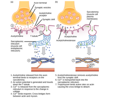
The neuromuscular junction is the synapse where a motor neuron communicates with a skeletal muscle fiber, triggering contraction.
Motor Neuron: Stimulates muscle fibers via axon branches.
Synaptic Vesicles: Contain acetylcholine (ACh), a neurotransmitter.
ACh Release: ACh is secreted into the synaptic cleft and binds to ligand-gated Na+ channels on the motor end plate of the sarcolemma.
Action Potential Generation: Binding of ACh opens Na+ channels, depolarizing the sarcolemma and generating an action potential.
Excitation-Contraction Coupling
Excitation-contraction coupling links the electrical signal from the neuron to the mechanical contraction of the muscle fiber.
Action Potential Transmission: The action potential travels along the sarcolemma and down T-tubules.
Calcium Release: Depolarization of T-tubules opens voltage-gated Ca2+ channels in the terminal cisternae of the sarcoplasmic reticulum (SR).
Troponin Activation: Ca2+ binds to troponin, causing tropomyosin to move and expose actin's active sites.
Crossbridge Formation: Myosin heads bind to actin, initiating contraction.
Muscle Relaxation: Occurs when ACh is degraded, Ca2+ is pumped back into the SR, and troponin/tropomyosin block actin sites.
Crossbridge Cycle and Muscle Contraction
The crossbridge cycle describes the repeated interactions between myosin and actin that drive muscle contraction.
ATP Hydrolysis: ATP is hydrolyzed, "cocking" the myosin head.
Crossbridge Formation: Myosin binds to actin's exposed active sites.
Power Stroke: Myosin releases ADP and Pi, pulling actin toward the M line.
Crossbridge Detachment: A new ATP binds to myosin, breaking the crossbridge.
Cycle Repeats: As long as Ca2+ and ATP are available, the cycle continues.
Muscle Relaxation
Muscle relaxation is achieved by removing the stimulus and restoring ionic conditions.
Acetylcholinesterase: Degrades ACh in the synaptic cleft.
Resting Potential: Sarcolemma returns to resting potential.
Calcium Reuptake: Ca2+ is pumped back into the SR.
Troponin/Tropomyosin: Block actin's active sites, preventing contraction.
Energy Requirements and Sources for Muscle Contraction
Muscle fibers require substantial ATP for contraction, ion pumping, and relaxation. Multiple mechanisms supply ATP:
Immediate Source: Creatine phosphate (CP) rapidly donates phosphate to ADP, forming ATP (effective for ~10 seconds).
Anaerobic Glycolysis: Glucose is converted to pyruvate and lactic acid, yielding 2 ATP per glucose (effective for ~1 minute).
Aerobic Catabolism: In the presence of oxygen, glucose, fatty acids, and amino acids are metabolized via the Krebs cycle and oxidative phosphorylation, yielding up to 36 ATP per glucose.
Formula:
Key Concepts Table
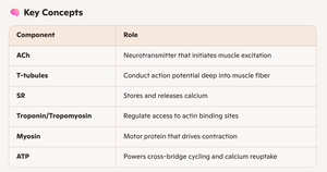
The following table summarizes the roles of major components involved in muscle contraction:
Component | Role |
|---|---|
ACh | Neurotransmitter that initiates muscle excitation |
T-tubules | Conduct action potential deep into muscle fiber |
SR | Stores and releases calcium |
Troponin/Tropomyosin | Regulate access to actin binding sites |
Myosin | Motor protein that drives contraction |
ATP | Powers cross-bridge cycling and calcium reuptake |
Example: During a sprint, muscle cells first use creatine phosphate for immediate energy, then switch to anaerobic glycolysis, and finally rely on aerobic metabolism for sustained activity.
Additional info: The notes expand on the original content by providing definitions, formulas, and examples for clarity and completeness.
