 뒤로
oct 19 (muscle)
스터디 가이드 - 스마트 노트
 자료에 맞춘 맞춤형 노트, 핵심 정의, 예시, 맥락을 확장해 제공합니다.
Muscle Tissue Overview
Introduction to Muscle Types
Muscle tissue is specialized for generating force and movement in response to physiological stimuli. There are three primary types of muscle tissue in the human body: skeletal muscle, cardiac muscle, and smooth muscle. Each type has distinct structural and functional characteristics.
Skeletal muscle: Voluntary, striated, multinucleated; responsible for body movement, posture, and heat production.
Cardiac muscle: Involuntary, striated, uninucleated; found only in the heart, responsible for pumping blood.
Smooth muscle: Involuntary, non-striated, uninucleated; found in walls of hollow organs, regulates passage of substances.
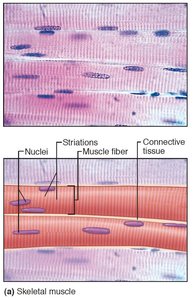
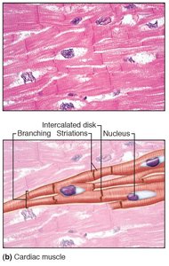
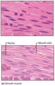
Skeletal Muscle Structure
Macroscopic Anatomy
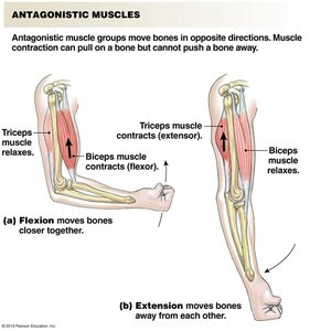
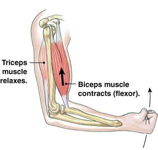
Skeletal muscles are usually attached to bones by tendons and are organized into antagonistic groups (flexors and extensors). The origin is the attachment closest to the trunk or stationary bone, while the insertion is more distal or mobile. Flexors bring bones together; extensors move bones apart.
Antagonistic muscle groups: Muscles that move bones in opposite directions.
Flexor: Contracts to bring bones closer together.
Extensor: Contracts to move bones away from each other.
Microscopic Anatomy
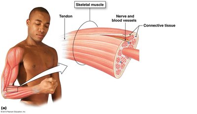
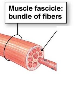
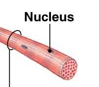
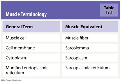
Skeletal muscle is composed of bundles of muscle fibers (cells), which are grouped into fascicles. Each muscle fiber is surrounded by a cell membrane called the sarcolemma and contains cytoplasm known as sarcoplasm. The modified endoplasmic reticulum is called the sarcoplasmic reticulum.
Muscle fiber: Single muscle cell, multinucleated, extends the length of the muscle.
Fascicle: Bundle of muscle fibers.
Connective tissue: Surrounds and supports muscle fibers and fascicles.
Muscle Terminology Table
The following table summarizes key terminology used in muscle anatomy:
General Term | Muscle Equivalent |
|---|---|
Muscle cell | Muscle fiber |
Cell membrane | Sarcolemma |
Cytoplasm | Sarcoplasm |
Modified endoplasmic reticulum | Sarcoplasmic reticulum |
Myofibrils and Sarcomere Structure
Myofibrils
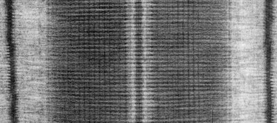
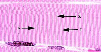
Each muscle fiber contains hundreds to thousands of myofibrils, which are the contractile units of the cell. Myofibrils are composed of repeating units called sarcomeres, which are responsible for the striated appearance of skeletal and cardiac muscle.
Striations: Result from the ordered arrangement of thick (myosin) and thin (actin) filaments.
Sarcomere: The basic functional unit of muscle contraction.
Sarcomere Model
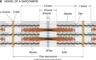
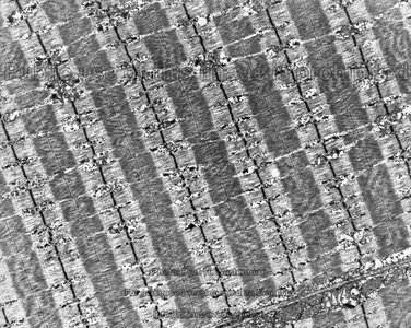
The sarcomere is defined by Z disks and contains several distinct regions:
Z disk: Attachment site for thin filaments (actin).
I band: Light band, contains only thin filaments.
A band: Dark band, contains thick filaments (myosin) and overlapping thin filaments.
H zone: Central region of A band, contains only thick filaments.
M line: Attachment site for thick filaments.
Titin and Nebulin: Structural proteins that stabilize and regulate filament length.
Myofilaments: Thin and Thick Filaments
Thin Filaments
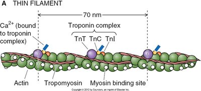
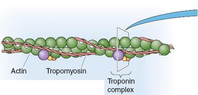
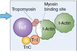
Thin filaments are primarily composed of F-actin, which forms a double-stranded helical polymer. Associated proteins include tropomyosin and the troponin complex (TnT, TnC, TnI), which regulate muscle contraction by controlling the binding of myosin to actin.
F-actin: Backbone of thin filaments, contains myosin binding sites.
Tropomyosin: Regulates access to myosin binding sites on actin.
Troponin complex:
TnT: Binds tropomyosin
TnC: Binds Ca2+
TnI: Inhibits contraction by binding actin
Thick Filaments
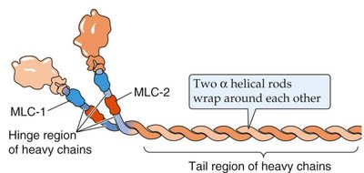
Thick filaments are composed of bundles of myosin molecules. Each myosin molecule has a head region that binds actin and hydrolyzes ATP, and a tail region formed by two intertwined heavy chains. Regulatory and essential light chains modulate ATPase activity and stabilize the myosin head.
Myosin head: Binds actin and ATP, responsible for force generation.
Regulatory light chain: Regulates ATPase activity.
Essential light chain: Stabilizes myosin head.
Muscle Contraction: The Sliding Filament Model
Mechanism of Contraction
Muscle contraction occurs via the sliding filament model, where myosin heads bind to actin and pull the thin filaments toward the center of the sarcomere, shortening the muscle. The force generated is referred to as muscle tension.
ATP: Provides energy for contraction.
Calcium ions (Ca2+): Trigger contraction by binding to troponin C.
Initiation of Skeletal Muscle Contraction
Neuromuscular Junction and Excitation-Contraction Coupling
The neuromuscular junction is the synaptic contact between a somatic motor neuron and a muscle fiber. An action potential in the motor neuron triggers the release of neurotransmitter, leading to an action potential in the muscle fiber and an increase in intracellular Ca2+, which initiates contraction.
Motor unit: A single motor neuron and all the muscle fibers it innervates.
Excitation-contraction coupling: Process linking action potential to muscle contraction.
Summary Table: Muscle Types
Muscle Type | Control | Striations | Nuclei | Main Function |
|---|---|---|---|---|
Skeletal | Voluntary | Present | Multinucleated | Movement, posture |
Cardiac | Involuntary | Present | Uninucleated | Pumping blood |
Smooth | Involuntary | Absent | Uninucleated | Regulation of organ function |
Key Terms and Concepts
Sarcolemma: Muscle cell membrane
Sarcoplasm: Muscle cell cytoplasm
Sarcoplasmic reticulum: Modified endoplasmic reticulum for Ca2+ storage
Myofibril: Contractile unit within muscle fiber
Sarcomere: Functional unit of contraction
Motor unit: Motor neuron and its innervated muscle fibers
Relevant Equations
Muscle force generation is dependent on ATP hydrolysis:
Calcium binding to troponin C initiates contraction:
*Additional info: Academic context was added to clarify muscle structure, function, and terminology, and to provide a self-contained study guide suitable for exam preparation.*
