 뒤로
Muscle Tissue: Structure, Function, and Contraction Mechanisms
스터디 가이드 - 스마트 노트
 자료에 맞춘 맞춤형 노트, 핵심 정의, 예시, 맥락을 확장해 제공합니다.
Muscle Tissue Types
Overview of Muscle Tissue
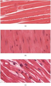
Muscle tissue is specialized for contraction and movement. There are three primary types of muscle tissue in the human body, each with distinct structural and functional characteristics.
Skeletal muscle: Voluntary, striated muscle responsible for movement and posture.
Cardiac muscle: Involuntary, striated muscle found only in the heart.
Smooth muscle: Involuntary, non-striated muscle found in walls of internal organs.
Skeletal Muscle Structure
Organization of Skeletal Muscle
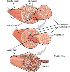
Skeletal muscle is organized into hierarchical structures, each surrounded by connective tissue layers that provide support and facilitate function.
Epimysium: The outermost layer of dense collagen fibers that surrounds the entire muscle, separating it from surrounding tissues.
Perimysium: Divides the muscle into compartments called fascicles, each containing bundles of muscle fibers.
Endomysium: Encases individual muscle fibers within a fascicle; contains capillaries and satellite cells for repair.
Muscle Fiber Anatomy
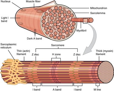
Each muscle fiber is a single, multinucleate cell containing specialized structures for contraction.
Sarcolemma: The plasma membrane of the muscle fiber.
Sarcoplasm: The cytoplasm of the muscle fiber.
Sarcoplasmic reticulum (SR): Specialized smooth endoplasmic reticulum for calcium storage.
Myofibrils: Cylindrical structures composed of myofilaments (actin and myosin).
Functional Units of Muscle Contraction
Sarcomere Structure
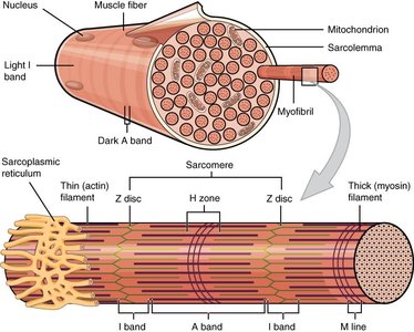
The sarcomere is the fundamental contractile unit of skeletal muscle, composed of organized arrangements of thick (myosin) and thin (actin) filaments.
Myofilaments: Protein filaments (actin and myosin) responsible for contraction.
Regulatory proteins: Troponin and tropomyosin control access to actin's active sites.
Striations: The banded appearance of skeletal muscle is due to the arrangement of actin and myosin filaments.
Neuromuscular Junction and Excitation
Neuromuscular Junction (NMJ)
The NMJ is the site where a motor neuron communicates with a muscle fiber, initiating contraction through neurotransmitter release.
Motor neuron: Innervates each muscle fiber at the NMJ.
Acetylcholine (ACh): Neurotransmitter released from synaptic vesicles, binds to receptors on the motor end-plate.
Action potential: Electrical signal generated by Na+ influx, spreads along the sarcolemma.
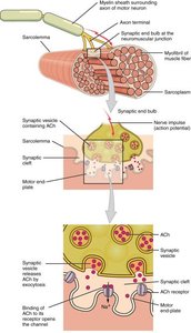
Excitation-Contraction Coupling
Mechanism of Muscle Contraction
Excitation-contraction coupling links the electrical excitation of the muscle fiber to the mechanical contraction process.
T-tubules: Carry the action potential into the interior of the cell.
Sarcoplasmic reticulum: Releases Ca2+ ions in response to the action potential.
Calcium ions: Bind to troponin, causing tropomyosin to move and expose actin's active sites.
Cross-Bridge Cycle
The cross-bridge cycle describes the interaction between actin and myosin during muscle contraction.
Myosin heads, cocked by ATP hydrolysis, bind to actin's active sites, forming cross-bridges.
ADP and Pi are released, and the myosin head pivots (power stroke), pulling actin filaments toward the center of the sarcomere.
ATP binds to myosin, causing it to detach from actin; ATP hydrolysis recocks the myosin head.
As long as Ca2+ and ATP are available, the cycle repeats.
Energy for Muscle Contraction
ATP Generation Pathways
Muscle contraction requires ATP, which is generated through several metabolic pathways.
Creatine phosphate: Provides rapid ATP regeneration for short bursts (about 15 seconds).
Glycolysis: Anaerobic breakdown of glucose to produce ATP and pyruvate.
Aerobic respiration: Occurs in mitochondria, producing large amounts of ATP from pyruvate when oxygen is available.
Summary Table: ATP Generation Pathways
Pathway | Duration | ATP Yield | Key Features |
|---|---|---|---|
Creatine Phosphate | ~15 seconds | 1 ATP per creatine phosphate | Rapid, short-term energy |
Glycolysis | ~1-2 minutes | 2 ATP per glucose | Anaerobic, produces pyruvate |
Aerobic Respiration | Long-term | ~34 ATP per glucose | Requires oxygen, efficient |
Muscle Contraction and Relaxation
Sliding Filament Model
Muscle contraction occurs as thin filaments slide past thick filaments, shortening the sarcomere and the muscle fiber.
Power stroke: Myosin heads pull actin filaments toward the center of the sarcomere.
Relaxation: Requires ATP to pump Ca2+ back into the SR and detach myosin from actin.
Role of Oxygen and Lactic Acid
During intense exercise, when oxygen is limited, glycolysis continues to provide ATP, and pyruvate is converted to lactic acid, which is transported to the liver for conversion back to glucose (Cori Cycle).
Lactic acid: Byproduct of anaerobic glycolysis; can cause muscle fatigue.
Cori Cycle: Process by which lactic acid is converted to glucose in the liver.
Key Terms and Definitions
Fascicle: Bundle of muscle fibers within a muscle.
Satellite cell: Stem cell involved in muscle repair.
Sarcolemma: Muscle cell membrane.
Sarcoplasm: Muscle cell cytoplasm.
Myofibril: Contractile organelle within muscle fiber.
Sarcomere: Functional unit of muscle contraction.
Troponin/Tropomyosin: Regulatory proteins controlling actin-myosin interaction.
Acetylcholine (ACh): Neurotransmitter at NMJ.
Creatine kinase: Enzyme facilitating ATP regeneration from creatine phosphate.
Important Equations
ATP Hydrolysis
ATP is hydrolyzed to provide energy for muscle contraction:
Creatine Phosphate Reaction
Creatine phosphate regenerates ATP:
Glycolysis
Glucose breakdown in glycolysis:
Aerobic Respiration
Pyruvate oxidation in mitochondria:
Summary Table: Muscle Tissue Types
Type | Location | Control | Appearance |
|---|---|---|---|
Skeletal | Attached to bones | Voluntary | Striated |
Cardiac | Heart | Involuntary | Striated |
Smooth | Walls of organs | Involuntary | Non-striated |
Example: Skeletal muscle contraction allows voluntary movement such as walking, while cardiac muscle contraction pumps blood throughout the body.
Additional info: Expanded explanations and tables were added for clarity and completeness based on academic context.
