 뒤로
Muscle Tissue: Structure, Function, and Mechanisms of Contraction
스터디 가이드 - 스마트 노트
 자료에 맞춘 맞춤형 노트, 핵심 정의, 예시, 맥락을 확장해 제공합니다.
Muscle Tissue Overview
Types of Muscle Tissue
Muscle tissue is a primary tissue in the human body, specialized for contraction and movement. There are three main types:
Skeletal muscle: Moves the body by pulling on bones; voluntary control.
Cardiac muscle: Found only in the heart; controls rhythmic contractions.
Smooth muscle: Controls movements inside the body, such as in blood vessels and organs.
Common Properties of Muscle Tissue
Excitability: Ability to respond to stimuli.
Contractility: Ability to shorten and generate force.
Extensibility: Ability to stretch without damage.
Elasticity: Ability to return to original shape after stretching.
Functions of Skeletal Muscle
Producing movement
Maintaining posture and body position
Supporting soft tissues
Guarding body entrances and exits
Maintaining body temperature
Storing nutrients
Organization of Skeletal Muscle
Muscle Structure at the Tissue Level
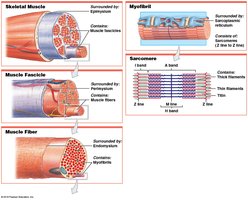
Skeletal muscles are composed of muscle tissue, connective tissues, blood vessels, and nerves. The organization is hierarchical:
Epimysium: Surrounds the entire muscle; connected to deep fascia.
Perimysium: Surrounds bundles of muscle fibers (fascicles); contains blood vessels and nerves.
Endomysium: Surrounds individual muscle fibers; contains capillaries, myosatellite cells, and nerve fibers.
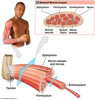
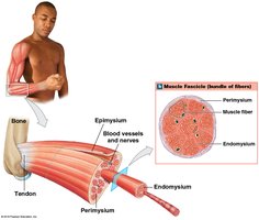
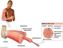
Collagen fibers from these layers converge to form tendons (bundles) or aponeuroses (sheets), which attach muscles to bones.
Skeletal Muscle Fibers
Characteristics and Development
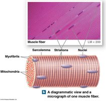
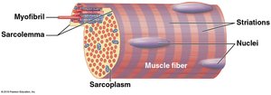
Skeletal muscle fibers are large, multinucleate cells formed by the fusion of embryonic myoblasts. They are also known as striated muscle cells due to their banded appearance.
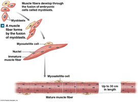
Internal Structure
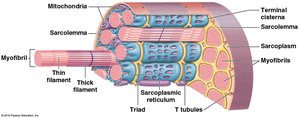
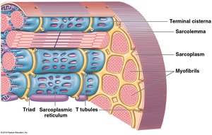
Sarcolemma: Plasma membrane surrounding the fiber.
Sarcoplasm: Cytoplasm of the muscle fiber.
Transverse tubules (T tubules): Invaginations that transmit action potentials into the cell.
Sarcoplasmic reticulum (SR): Specialized for calcium storage and release; forms terminal cisternae and triads with T tubules.
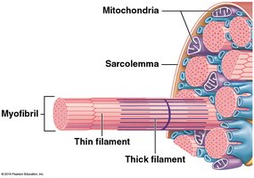
Myofibrils: Bundles of protein filaments responsible for contraction; contain thin (actin) and thick (myosin) filaments.
Sarcomere Structure
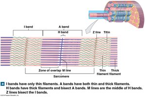
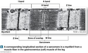
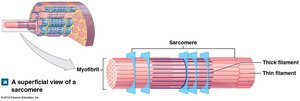
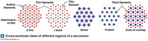
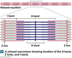
The sarcomere is the smallest functional unit of a muscle fiber. Its arrangement of thick and thin filaments produces the striated pattern.
A band: Dark band; contains thick filaments.
I band: Light band; contains thin filaments.
H band: Area with only thick filaments.
M line: Center of A band; stabilizes thick filaments.
Z line: Boundary between sarcomeres; bisects I bands.
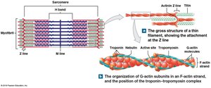
Titin: Elastic protein maintaining alignment and aiding in recoil.
Thin and Thick Filaments
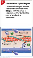
Thin filaments: Composed of F-actin (two rows of G-actin), nebulin, tropomyosin (covers active sites), and troponin (binds Ca2+).
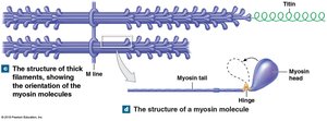
Thick filaments: Composed of myosin molecules (head and tail regions); core of titin for elasticity.
Sliding-Filament Theory
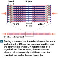
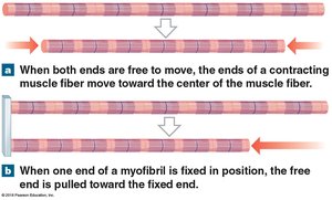
Muscle contraction occurs as thin filaments slide toward the center of the sarcomere, causing H and I bands to narrow, Z lines to move closer, and the A band to remain constant.
The Neuromuscular Junction
Components and Events
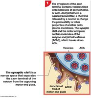
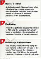
The neuromuscular junction (NMJ) is the synapse between a motor neuron and a skeletal muscle fiber. The process of muscle contraction is initiated by neural stimulation:
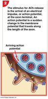
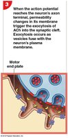
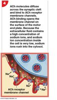
Motor neuron releases acetylcholine (ACh) into the synaptic cleft.
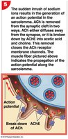
ACh binds to Na+ channels on the muscle fiber, causing depolarization.
An action potential is generated and travels along the sarcolemma and T tubules.
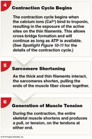
Ca2+ is released from the SR, binds to troponin, and exposes active sites on actin.
The contraction cycle begins: cross-bridge formation, power stroke, detachment, and reactivation.
Muscle Tension Production
Mechanisms of Tension
The amount of tension produced by a muscle fiber depends on:
Number of power strokes performed
Resting length of the fiber at stimulation
Frequency of stimulation
Length–Tension Relationship
Maximum tension is produced when the maximum number of cross-bridges is formed, which occurs when the zone of overlap between thick and thin filaments is optimal.
Frequency of Stimulation
A single neural stimulation produces a twitch.
Sustained contractions require repeated stimuli.
A myogram shows tension development over time.
Treppe and Wave Summation
Treppe: Stair-step increase in tension with repeated stimulation after relaxation.
Wave summation: Increased tension due to repeated stimulation before relaxation ends.
Tetanus
Incomplete tetanus: Near-maximum tension with rapid cycles of contraction and relaxation.
Complete tetanus: Continuous contraction with no relaxation phase; all cross-bridges formed.
Types of Muscle Contractions
Motor Units and Recruitment
A motor unit consists of a motor neuron and all the muscle fibers it controls. Recruitment increases the number of active motor units, producing a smooth increase in tension.
Muscle Tone
Normal tension and firmness at rest
Stabilizes bones and joints
Maintains posture
Classification of Contractions
Isotonic contractions: Muscle changes length (concentric: shortens; eccentric: lengthens).
Isometric contractions: Muscle develops tension without changing length.
Load and Speed of Contraction
The heavier the load, the slower the contraction and the less the muscle shortens.
Energy for Muscle Contraction
ATP and Metabolic Pathways
ATP is the direct energy source for muscle contraction. Muscles store limited ATP and must regenerate it through:
Direct phosphorylation by creatine phosphate (CP)
Anaerobic metabolism (glycolysis)
Aerobic metabolism (citric acid cycle and electron transport chain)
Muscle Metabolism
At rest: Metabolize fatty acids, store glycogen and CP
Moderate activity: Aerobic breakdown of glucose
Peak activity: Glycolysis produces lactate
Recovery and Oxygen Debt
Recovery period: Time required for muscles to return to normal after exertion.
Cori cycle: Lactate is transferred to the liver, converted to pyruvate, then glucose.
Oxygen debt (EPOC): Increased oxygen demand after exercise to restore metabolic balance.
Heat Production and Hormonal Effects
Active muscles produce heat, contributing to body temperature regulation.
Hormones (growth hormone, testosterone, thyroid hormones, epinephrine) increase muscle metabolism.
Muscle Performance and Fiber Types
Types of Muscle Fibers
Fast fibers: Contract quickly, large diameter, few mitochondria, fatigue rapidly.
Slow fibers: Contract slowly, small diameter, many mitochondria, high oxygen supply, contain myoglobin.
Intermediate fibers: Mid-sized, little myoglobin, slower to fatigue than fast fibers.
Muscle Hypertrophy and Atrophy
Hypertrophy: Increase in muscle size, myofibrils, mitochondria, and glycogen reserves due to training.
Atrophy: Reduction in muscle size, tone, and power due to inactivity.
Aging and Muscle Fatigue
Muscle fibers decrease in diameter and elasticity with age.
Fibrosis increases, exercise tolerance and recovery decrease.
Muscle fatigue: Inability to sustain performance due to metabolic depletion, damage, and pH changes.
Physical Conditioning
Anaerobic endurance: Uses fast fibers, stimulates hypertrophy; improved by brief, intensive workouts.
Aerobic endurance: Supported by mitochondria; improved by sustained, low-intensity activity.
Cardiac and Smooth Muscle Tissue
Cardiac Muscle Tissue
Found only in the heart; striated, small, branched cells with single nucleus.
Short, wide T tubules; no triads; SR lacks terminal cisternae.
Dependent on aerobic metabolism; contains myoglobin and many mitochondria.
Cells connected by intercalated discs (gap junctions and desmosomes).
Automaticity: Contraction without neural stimulation, controlled by pacemaker cells.
Smooth Muscle Tissue
Long, slender, spindle-shaped cells with single central nucleus.
No T tubules, myofibrils, or sarcomeres; nonstriated.
Scattered thick filaments with many myosin heads; thin filaments attached to dense bodies.
Dense bodies connect cells and transmit contractions; no tendons or aponeuroses.
Functional Characteristics of Smooth Muscle
Excitation–contraction coupling: Ca2+ binds to calmodulin, activating myosin light chain kinase.
Length–tension relationship: Can contract even when stretched; plasticity allows function over wide length range.
Control of contractions: Multiunit (innervated) and visceral (not innervated, controlled by pacesetter cells).
Smooth muscle tone: Normal background activity, modulated by neural, hormonal, or chemical factors.
