 뒤로
Muscle Tissue: Structure, Function, and Physiology
스터디 가이드 - 스마트 노트
 자료에 맞춘 맞춤형 노트, 핵심 정의, 예시, 맥락을 확장해 제공합니다.
Muscle Tissue
Overview of Muscle Tissue
Muscle tissue is essential for movement, posture, and various involuntary processes in the body. There are three main types of muscle tissue: skeletal, cardiac, and smooth, each with distinct structures and functions.
Skeletal muscle: Attached to bones, responsible for voluntary movements.
Cardiac muscle: Found in the heart, responsible for pumping blood.
Smooth muscle: Located in walls of hollow organs, responsible for involuntary movements.
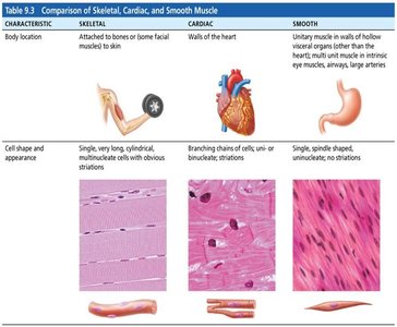
Types of Muscle Tissue
Comparison of Skeletal, Cardiac, and Smooth Muscle
Each muscle type has unique characteristics regarding location, structure, and control.
Characteristic | Skeletal | Cardiac | Smooth |
|---|---|---|---|
Body Location | Attached to bones or skin | Walls of the heart | Walls of hollow organs (e.g., stomach, arteries) |
Cell Shape & Appearance | Long, cylindrical, multinucleate, striated | Branching chains, uni- or binucleate, striated | Spindle-shaped, uninucleate, non-striated |
Control | Voluntary | Involuntary | Involuntary |
Connective Tissue Components of Skeletal Muscle
Organization and Structure
Skeletal muscle is organized into bundles and surrounded by connective tissue sheaths that provide support and transmit force.
Epimysium: Surrounds the entire muscle.
Perimysium: Surrounds bundles of muscle fibers (fascicles).
Endomysium: Surrounds individual muscle fibers.
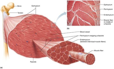
Structure of Skeletal Muscle Fibers
Muscle Fiber Anatomy
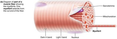
Skeletal muscle fibers are long, multinucleated cells containing specialized organelles for contraction.
Sarcolemma: Plasma membrane of the muscle fiber.
Sarcoplasm: Cytoplasm containing myofibrils, glycosomes, and myoglobin.
Myofibrils: Rodlike structures responsible for muscle contraction, composed of repeating units called sarcomeres.
Sarcomere Structure
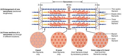
The sarcomere is the fundamental contractile unit of muscle, defined by the area between two Z discs. It contains thick (myosin) and thin (actin) filaments arranged in a precise pattern, producing the striated appearance of skeletal and cardiac muscle.
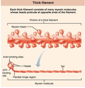
Thick filaments: Composed of myosin molecules.
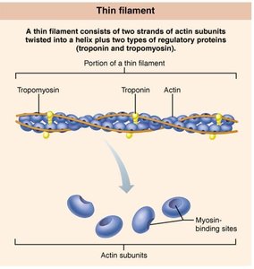
Thin filaments: Composed of actin, tropomyosin, and troponin.
Elastic filaments: Composed of titin, providing elasticity and stability.
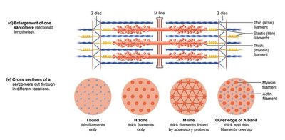
Myofilament Details
Thick and thin filaments interact to produce muscle contraction. Myosin heads on thick filaments bind to actin on thin filaments, forming cross-bridges.
Myosin: Has heads with ATPase activity and actin-binding sites.
Actin: Contains myosin-binding sites, regulated by tropomyosin and troponin.
Functional Organization of Muscle
Levels of Organization
Muscle tissue is organized hierarchically from the whole muscle down to the molecular level.
Muscle: Composed of fascicles.
Fascicle: Bundle of muscle fibers.
Muscle fiber: Single muscle cell containing myofibrils.
Myofibril: Composed of repeating sarcomeres.
Sarcomere: Contains thick and thin filaments.
Events of Muscle Contraction and Relaxation
Excitation-Contraction Coupling
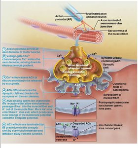
Muscle contraction is initiated by a nerve impulse that leads to the release of calcium ions and the interaction of actin and myosin.
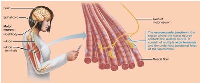
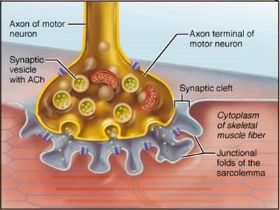
Neuromuscular junction: Site where a motor neuron stimulates a muscle fiber.
Acetylcholine (ACh): Neurotransmitter that triggers muscle action potential.
Action potential: Electrical signal that travels along the sarcolemma and T tubules, leading to calcium release from the sarcoplasmic reticulum.
Action Potential and Muscle Fiber Response
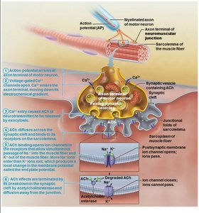
The action potential in the muscle fiber leads to depolarization and repolarization, which are essential for muscle contraction and relaxation.
Depolarization: Na+ channels open, Na+ enters the cell, making the membrane potential more positive.
Repolarization: K+ channels open, K+ exits the cell, restoring the resting membrane potential.
Energy Sources for Muscle Contraction
ATP Regeneration Pathways
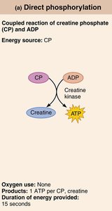
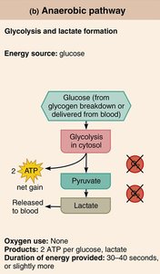
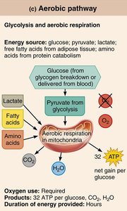
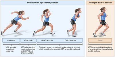
ATP is the immediate source of energy for muscle contraction. Because ATP stores are limited, muscle fibers regenerate ATP through three main pathways:
Direct phosphorylation of ADP by creatine phosphate (CP): Fastest, short duration.
Anaerobic pathway (glycolysis and lactic acid formation): Moderate speed, produces ATP without oxygen.
Aerobic pathway (aerobic respiration): Slowest, but provides the most ATP for prolonged activity.
Principles of Muscle Contraction
All-or-None Principle
The all-or-none principle states that a muscle fiber contracts fully when stimulated by a threshold stimulus; if the stimulus is below threshold, no contraction occurs. The strength of contraction is not graded at the level of the individual fiber.
Action potential: Always the same size for a given fiber; does not vary in strength.
Implication: Ensures reliable transmission of signals and prevents loss of information.
Types of Skeletal Muscle Contraction
Muscle contractions can be classified based on whether the muscle changes length during contraction.
Isometric contraction: Muscle tension increases, but muscle does not change length (e.g., holding a weight steady).
Isotonic contraction: Muscle changes length to move a load.
Concentric contraction: Muscle shortens while generating force (e.g., lifting a book).
Eccentric contraction: Muscle lengthens while maintaining force (e.g., lowering a book).
Clinical Implications
Muscle Disorders
Duchenne muscular dystrophy (DMD): Genetic disorder causing progressive muscle weakness due to defective dystrophin protein.
Myasthenia gravis: Autoimmune disease leading to muscle weakness by reducing the number of functional ACh receptors.
Rigor mortis: Postmortem stiffening of muscles due to lack of ATP, preventing detachment of myosin from actin.
Disuse atrophy: Muscle wasting due to inactivity or loss of neural stimulation.
