 뒤로
Muscle Tissue: Structure, Function, and Physiology
스터디 가이드 - 스마트 노트
 자료에 맞춘 맞춤형 노트, 핵심 정의, 예시, 맥락을 확장해 제공합니다.
Muscle Tissue
I. Characteristics of Muscle Tissue
Muscle tissue is specialized for contraction and is essential for movement and force generation in the body. It possesses several unique physiological properties:
Excitability: The ability to receive and respond to stimuli, typically from the nervous system.
Contractility: The ability to shorten forcibly when stimulated, generating tension.
Extensibility: The ability to be stretched or extended without being damaged.
Elasticity: The ability to return to its original length after contraction or extension.
II. Functions of Muscle Tissue
Muscle tissue performs several vital functions in the human body:
Movement: Muscles produce movement of the body and its parts by contracting and pulling on bones or skin.
Maintenance of posture: Continuous muscle contractions help maintain body posture and position.
Stabilization of joints: Muscles reinforce and stabilize joints.
Heat generation: Muscle contractions produce heat, which is important for maintaining body temperature.
Protection of internal organs: Some muscles provide protection to internal organs by forming muscular walls.
III. Types of Muscle Tissue
There are three main types of muscle tissue, each with distinct structure and function:
Skeletal Muscle: Voluntary, striated muscle attached to bones; responsible for body movement.
Cardiac Muscle: Involuntary, striated muscle found only in the heart; responsible for pumping blood.
Smooth Muscle: Involuntary, non-striated muscle found in walls of hollow organs (e.g., intestines, blood vessels).
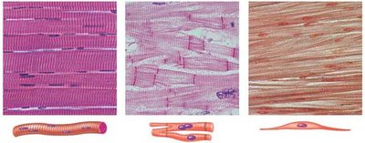
IV. Skeletal Muscle – Gross Anatomy (Organ Level)
Skeletal muscle is an organ composed of muscle fibers, connective tissue, blood vessels, and nerves. Its structure is organized into several layers of connective tissue sheaths:
Epimysium: Dense irregular connective tissue that surrounds the entire muscle, bundling fascicles together.
Perimysium: Fibrous connective tissue that wraps around each fascicle (a bundle of muscle fibers).
Endomysium: Delicate connective tissue that surrounds each individual muscle fiber (cell).
These connective tissue sheaths are continuous with each other and with tendons, which attach muscle to bone.
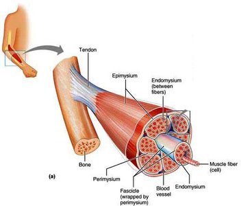
V. Skeletal Muscle – Microscopic Anatomy (Cellular Level)
A. Skeletal Muscle Cell (Muscle Fiber)
Skeletal muscle fibers are long, cylindrical cells formed by the fusion of hundreds of embryonic myoblasts. Some myoblasts remain as satellite cells, which aid in muscle repair and regeneration.
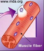
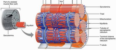
Sarcolemma: The plasma membrane of a muscle cell.
Sarcoplasm: The cytoplasm of a muscle cell, containing organelles, stored glycogen, myoglobin (an oxygen-binding pigment), myofibrils, and the sarcoplasmic reticulum (SR).
B. Myofibrils
Myofibrils are rodlike structures within muscle fibers, responsible for contraction. They contain contractile elements called myofilaments.
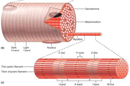
C. Myofilaments and Striations
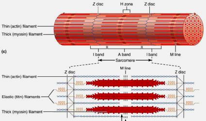
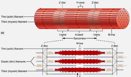
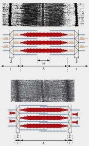
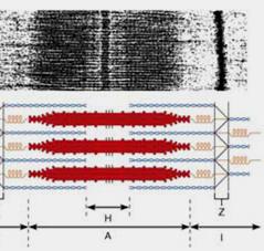
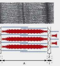
Myofilaments are organized into thick (myosin) and thin (actin) filaments, arranged in parallel and in register, producing the visible striations of skeletal muscle.
Dark bands (A bands): Regions with thick filaments (myosin).
Light bands (I bands): Regions with thin filaments (mostly actin).
M line: Middle of the A band, containing connecting proteins.
Z disc: Middle of the I band, anchoring thin filaments.
D. Sarcomeres
The sarcomere is the smallest contractile unit of muscle, defined as the segment between two Z discs. Sarcomeres are arranged end-to-end along the length of each myofibril.
Titin: An elastic protein that helps anchor and center thick filaments between Z discs.
Thick and thin filaments are interdigitated within the sarcomere.
E. Thick and Thin Filaments
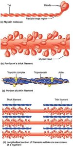
Thick Filament: Composed of bundled myosin protein molecules. Myosin heads protrude and bind to actin during contraction.
Thin Filament: Composed mainly of F-actin, with associated proteins tropomyosin (covers myosin binding sites) and troponin (binds Ca2+ and controls tropomyosin position).
F. Sarcoplasmic Reticulum and T-tubules
The sarcoplasmic reticulum (SR) is a specialized smooth endoplasmic reticulum that stores and releases Ca2+ for muscle contraction. T-tubules are invaginations of the sarcolemma that transmit action potentials deep into the muscle fiber. The triad consists of a T-tubule flanked by two terminal cisternae of the SR.
VI. Sliding Filament Theory of Contraction
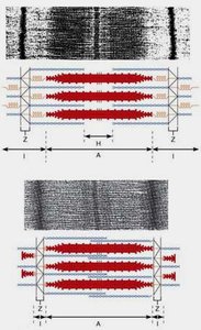
The sliding filament theory explains how muscles contract: thin filaments slide past thick filaments, increasing their overlap and shortening the sarcomere, which leads to muscle contraction.
VII. Physiology of Skeletal Muscle Fiber and Contraction
For a skeletal muscle fiber to contract, it must be stimulated by a nerve impulse. The process involves several steps:
Electrical impulse travels along the sarcolemma and through T-tubules.
This triggers the release of Ca2+ from the SR.
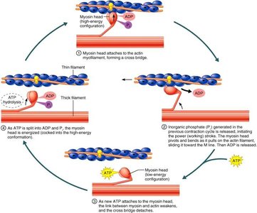
Myosin heads bind to actin, forming crossbridges.
Crossbridge cycling occurs, pulling actin filaments past myosin filaments.
A. Nerve Stimulus at the Neuromuscular Junction (NMJ)
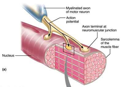
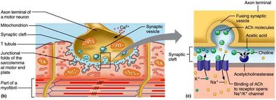
The NMJ is a specialized synapse where a motor neuron communicates with a muscle fiber. The axon terminal releases the neurotransmitter acetylcholine (ACh), which binds to receptors on the muscle fiber's motor endplate, initiating an action potential.
B. Generation and Propagation of Action Potential
Depolarization at the end plate (end plate potential) can trigger an action potential (AP) if it reaches threshold. The AP is a wave of depolarization that spreads along the sarcolemma, leading to muscle contraction. The AP is an all-or-none event and lasts about 1-2 milliseconds.
C. Excitation-Contraction Coupling (E-C Coupling)
Excitation-contraction coupling is the process by which an action potential triggers muscle contraction:
Neuron stimulation generates an AP in the muscle fiber.
The AP causes Ca2+ release from the SR.
Ca2+ activates crossbridge formation between myosin and actin.
Myosin pulls actin, shortening the sarcomere.
Ca2+ is reabsorbed into the SR, ending contraction.
Crossbridges detach, and the muscle relaxes.
D. Recovery after E-C Coupling
Clearance of ACh: Acetylcholine is removed from the synaptic cleft by reuptake, enzymatic breakdown by acetylcholinesterase, or diffusion away from the cleft.
Re-uptake of Ca2+: Ca2+ is rapidly pumped back into the SR, lowering cytosolic Ca2+ concentration and allowing muscle relaxation.
Table: Comparison of Muscle Tissue Types
Feature | Skeletal Muscle | Cardiac Muscle | Smooth Muscle |
|---|---|---|---|
Control | Voluntary | Involuntary | Involuntary |
Striations | Present | Present | Absent |
Location | Attached to bones | Heart | Walls of hollow organs |
Cell Shape | Long, cylindrical | Branched | Spindle-shaped |
Nuclei | Multiple, peripheral | Single, central | Single, central |
