 뒤로
Muscle Tissue: Structure, Function, and Physiology
스터디 가이드 - 스마트 노트
 자료에 맞춘 맞춤형 노트, 핵심 정의, 예시, 맥락을 확장해 제공합니다.
Muscle Tissue
Overview of Muscle Tissue
Muscle tissue is essential for movement, posture, and various physiological functions. It is classified into three main types: skeletal, cardiac, and smooth muscle, each with distinct structural and functional characteristics.
Contractility: Ability to shorten and produce movement.
Extensibility: Ability to stretch and return to resting length.
Excitability: Ability to respond to stimuli.
Highly vascularized: Ensures nourishment and waste removal.
Types of Muscle Tissue
Muscle tissue is categorized based on structure, function, and location.
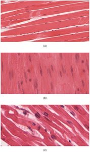
Skeletal Muscle: Multinucleated, striated, voluntary; attached to bones for movement.
Cardiac Muscle: 1-2 nuclei, striated, involuntary; found in the heart, responsible for pumping blood.
Smooth Muscle: Single nucleus, non-striated, involuntary; found in walls of organs and blood vessels.
Cardiac Muscle
Structure and Function
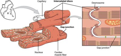
Cardiac muscle fibers are branched and interconnected by intercalated discs, which contain gap junctions and desmosomes. These structures allow for synchronized contraction and electrical coupling, forming a functional syncytium.
Intercalated discs: Specialized junctions for coordination.
Gap junctions: Allow rapid ion flow and depolarization.
Desmosomes: Anchor fibers during contraction.
Smooth Muscle
Structure and Function
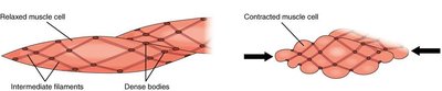
Smooth muscle cells are spindle-shaped, non-striated, and contain actin and myosin filaments anchored by dense bodies. Contraction is regulated by calcium binding to calmodulin, not troponin.
Dense bodies: Anchor thin filaments.
Calmodulin: Regulatory protein for contraction.
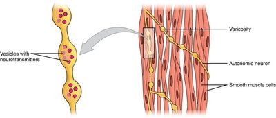
Varicosities: Sites of neurotransmitter release from autonomic neurons.
Skeletal Muscle
Structure and Organization
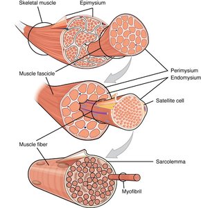
Skeletal muscle is organized into layers of connective tissue: epimysium, perimysium, and endomysium. Muscle fibers are multinucleated and contain myofibrils, which are composed of repeating units called sarcomeres.
Epimysium: Surrounds entire muscle.
Perimysium: Surrounds fascicles.
Endomysium: Surrounds individual fibers.
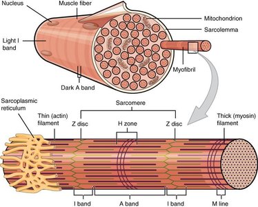
Myofibril: Cylindrical structure containing sarcomeres.
Sarcomere Structure
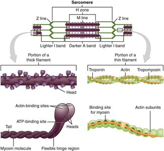
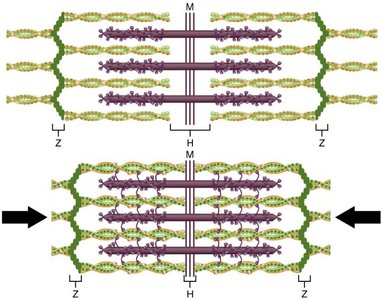
The sarcomere is the functional unit of skeletal muscle, defined by Z-lines. It contains thick (myosin) and thin (actin) filaments, which interact during contraction.
Z-line: Boundary of sarcomere.
I band: Light region with thin filaments.
A band: Dark region with thick filaments.
H zone: Central region with only thick filaments.
Regulatory proteins: Troponin and tropomyosin control access to myosin-binding sites.
Neuromuscular Junction and Motor Units
Neuromuscular Junction (NMJ)
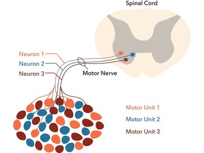
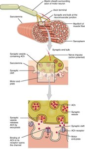
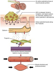
The NMJ is the site where a motor neuron communicates with a muscle fiber, initiating contraction via neurotransmitter release (acetylcholine).
Motor unit: A motor neuron and all the muscle fibers it innervates.
Recruitment: Activation of additional motor units increases contraction strength.
Muscle Contraction: Sliding Filament Model
Mechanism of Contraction
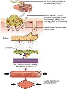
Muscle contraction occurs as myosin heads bind to actin, forming cross-bridges and pulling thin filaments toward the M line. This process requires calcium and ATP.
Cross-bridge cycling: Myosin heads attach, pivot, detach, and re-cock.
ATP: Required for detachment and re-cocking of myosin heads.
Calcium: Binds to troponin, exposing myosin-binding sites.
Muscle Relaxation
Mechanism of Relaxation
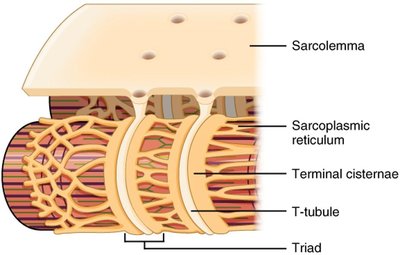
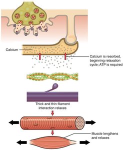
Relaxation occurs when nerve signals cease, calcium is pumped back into the sarcoplasmic reticulum, and ATP is used to reset the system. Tropomyosin covers myosin-binding sites, preventing further contraction.
ATP: Required for calcium reuptake and myosin head detachment.
Calcium: Released from troponin and pumped into SR.
Sources of ATP for Muscle Contraction
ATP Generation Pathways
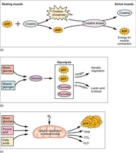
Muscle fibers rely on several mechanisms to generate ATP for contraction:
Creatine phosphate: Rapid, short-term energy source.
Glycolysis: Anaerobic breakdown of glucose for moderate energy.
Aerobic respiration: Efficient, long-term energy production using oxygen.
Muscle Attachments and Actions
Lever Systems and Muscle Roles
Skeletal muscles attach to bones via tendons and produce movement by acting as levers. Muscles are classified by their roles in movement: agonist, antagonist, synergist, and fixator.
Origin: Fixed attachment point.
Insertion: Movable attachment point.
Agonist: Prime mover.
Antagonist: Opposes the agonist.
Synergist: Assists the agonist.
Fixator: Stabilizes joints.
Summary Table: Muscle Types
Type | Structure | Function | Location |
|---|---|---|---|
Skeletal | Multinucleated, striated | Voluntary movement | Attached to bones |
Cardiac | 1-2 nuclei, striated, branched, intercalated discs | Pumps blood | Heart |
Smooth | Single nucleus, non-striated, spindle-shaped | Moves substances through organs | Walls of organs, blood vessels |
Key Equations
ATP Hydrolysis:
Creatine Phosphate Reaction:
Glycolysis:
Aerobic Respiration:
