 뒤로
Muscle Tissue: Structure, Function, and Physiology
스터디 가이드 - 스마트 노트
 자료에 맞춘 맞춤형 노트, 핵심 정의, 예시, 맥락을 확장해 제공합니다.
Muscle Tissue
Types and Functions of Muscle Tissue
Muscle tissue is a primary tissue responsible for contraction and movement in the human body. There are three main types of muscle tissue, each with distinct functions:
Skeletal muscle: Moves the body by pulling on bones.
Cardiac muscle: Pumps blood through the cardiovascular system.
Smooth muscle: Pushes fluids and solids through internal passageways and organs.
Common properties of muscle tissue include:
Excitability: Ability to receive and respond to stimuli.
Contractility: Ability of cells to shorten.
Extensibility: Ability of the muscle to stretch.
Elasticity: Ability of the muscle to recoil to its resting length.
Primary functions of skeletal muscle:
Producing movement
Maintaining posture and body position
Supporting soft tissues
Guarding body entrances and exits
Maintaining body temperature
Storing nutrients
Organization of Skeletal Muscle
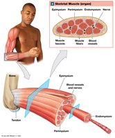
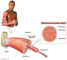
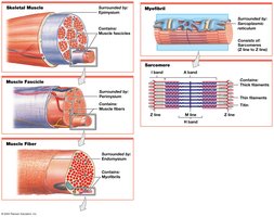
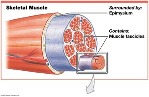
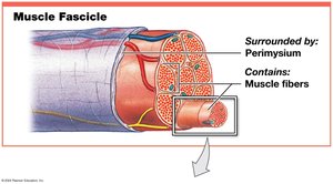
Skeletal muscles are composed of muscle tissue, connective tissues, blood vessels, and nerves. The organization at the tissue level involves three layers of connective tissue:
Epimysium: Surrounds the entire muscle and separates it from surrounding tissues.
Perimysium: Surrounds individual fascicles (bundles of muscle fibers).
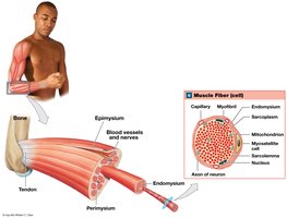
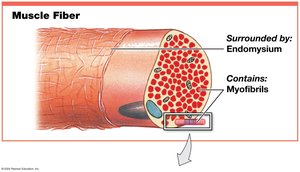
Endomysium: Surrounds individual muscle cells (muscle fibers).
The collagen fibers from these layers converge to form tendons or aponeuroses, which attach muscles to bones.
Skeletal Muscle Fibers and Sarcomere Structure
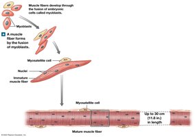
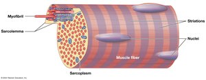
Skeletal muscle fibers are large, multinucleate cells formed by the fusion of myoblasts. They are striated due to the arrangement of myofibrils, which are collections of contractile myofilaments.
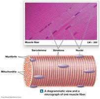
Key components:
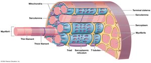
Sarcolemma: Plasma membrane of a muscle fiber.
Sarcoplasm: Cytoplasm of a muscle fiber.
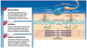
Transverse tubules (T tubules): Transmit action potentials into the cell interior.
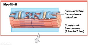
Sarcoplasmic reticulum (SR): Stores and releases calcium ions; forms terminal cisternae.
Myofibrils: Collections of myofilaments responsible for contraction.
Myofilaments: Thin (actin) and thick (myosin) filaments.
Sarcomere Structure
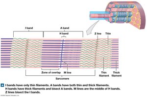
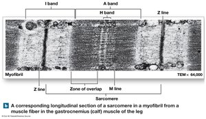
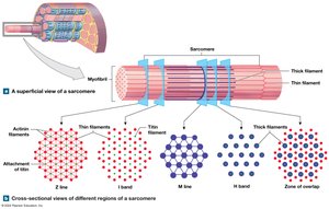
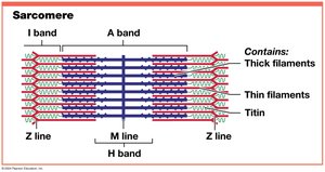
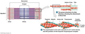
Sarcomeres are the smallest contractile units of muscle fibers, consisting of organized thin and thick filaments. Their arrangement produces visible striations.
A bands: Dark bands, length of thick filaments.
M line: Center of A band, stabilizes thick filaments.
H band: Only thick filaments.
Zone of overlap: Region where thick and thin filaments overlap.
I bands: Light bands, only thin filaments.
Z lines: Boundaries between sarcomeres.
Titin: Elastic protein, maintains alignment and restores length.
Thin and Thick Filaments
Thin filaments are primarily composed of actin, with associated proteins:
F-actin: Twisted strand of G-actin molecules.
Nebulin: Holds F-actin strands together.
Tropomyosin: Covers active sites on G-actin.
Troponin: Binds calcium ions and moves tropomyosin.
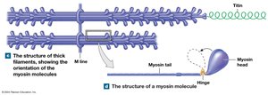
Thick filaments are composed of myosin molecules:
Myosin tail: Binds to other myosin molecules.
Myosin head: Projects toward nearest thin filament.
Titin: Core of thick filament, acts as a molecular spring.
Sliding-Filament Theory
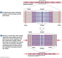
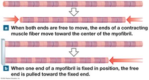
During contraction, thin filaments slide toward the M line alongside thick filaments, causing:
H and I bands to narrow
Zones of overlap to widen
Z lines to move closer together
A band width remains constant
Neuromuscular Junction and Muscle Contraction
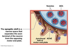
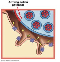
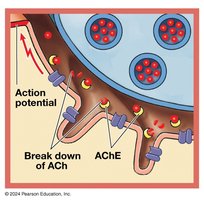
The neuromuscular junction (NMJ) is the synapse between a motor neuron and a skeletal muscle fiber. Muscle contraction is initiated by neural stimulation:
Axon terminal: End of motor neuron.
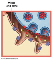
Motor end plate: Folded membrane of muscle fiber.
Synaptic cleft: Space between axon terminal and motor end plate.
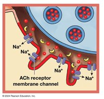
Acetylcholine (ACh): Neurotransmitter released into synaptic cleft.
ACh receptor: Opens Na+ channels, depolarizes motor end plate.
Excitation–Contraction Coupling
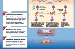
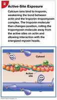
The action potential travels down T tubules, triggering calcium release from the SR. Calcium binds to troponin, moving tropomyosin and exposing actin active sites, allowing contraction to begin.
Contraction Cycle and Cross-Bridge Formation
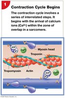
The contraction cycle involves cross-bridge formation, power stroke, and detachment, powered by ATP:
Myosin heads bind to actin (cross-bridge).
Power stroke: Myosin head pivots, pulling actin toward M line.
ATP binds to myosin, causing detachment and reactivation.
Muscle Tension and Types of Contraction
Tension is the pull force generated by muscle contraction. The amount of tension depends on:
Number of power strokes
Resting length of muscle fiber (length-tension relationship)
Frequency of stimulation
Length–Tension Relationship
Maximum tension is produced when the maximum number of cross-bridges is formed at the optimal sarcomere length.
Frequency of Stimulation
Twitch: Single stimulus-contraction-relaxation sequence.
Treppe: Increase in peak tension with repeated stimulations.
Wave summation: Increased tension due to successive stimuli.
Tetanus: Maximum tension, either incomplete or complete.
Types of Muscle Contraction
Isotonic contraction: Muscle changes length (concentric: shortens, eccentric: lengthens).
Isometric contraction: Muscle develops tension but does not change length.
Energy for Muscle Contraction
Muscle contraction requires ATP, which is generated by:
Creatine phosphate (CP): Stores energy for rapid ATP regeneration.
Glycolysis: Anaerobic breakdown of glucose.
Aerobic metabolism: Oxygen-dependent ATP production in mitochondria.
At rest, muscles use fatty acids; during activity, they use glucose and CP. At peak activity, glycolysis predominates, producing lactic acid and causing fatigue.
Muscle Performance and Fiber Types
Muscle performance depends on force and endurance, which are influenced by fiber type and conditioning:
Fast fibers: Quick, powerful contractions, fatigue rapidly.
Slow fibers: Slower, sustained contractions, high endurance.
Intermediate fibers: Characteristics between fast and slow fibers.
Cardiac and Smooth Muscle Tissue
Cardiac Muscle
Found only in the heart
Small, branched, striated cells with a single nucleus
Connected by intercalated discs
Automaticity: contracts without neural stimulation
Smooth Muscle
Found in internal organs and passageways
Spindle-shaped, non-striated cells with a single nucleus
Contractions controlled by neural, hormonal, or chemical factors
Plasticity: functions over a wide range of lengths
Example: Smooth muscle in the digestive tract moves food, while cardiac muscle pumps blood throughout the body.
Additional info: This summary covers the structure, function, and physiology of muscle tissue, including skeletal, cardiac, and smooth muscle, as well as mechanisms of contraction, energy metabolism, and muscle performance.
