 뒤로
Muscle Tissue: Structure, Function, and Physiology
스터디 가이드 - 스마트 노트
 자료에 맞춘 맞춤형 노트, 핵심 정의, 예시, 맥락을 확장해 제공합니다.
Muscle Tissue
Overview of Muscle Tissue
Muscle tissue is specialized for contraction and is responsible for movement of the body and its parts. There are three main types of muscle tissue in the human body: skeletal muscle, cardiac muscle, and smooth muscle. Each type has distinct structural and functional characteristics that suit its role in the body.

Types of Muscle Tissue
Skeletal Muscle: Voluntary, striated muscle attached to bones; responsible for body movement.
Cardiac Muscle: Involuntary, striated muscle found only in the heart; responsible for pumping blood.
Smooth Muscle: Involuntary, non-striated muscle found in walls of hollow organs (e.g., digestive tract, blood vessels).
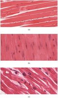
Example: Skeletal muscle enables athletes to perform rapid and powerful movements, while smooth muscle controls the movement of food through the digestive tract.
Structure of Skeletal Muscle
Connective Tissue Layers
Skeletal muscle is organized into bundles of muscle fibers called fascicles. Each muscle fiber is surrounded by connective tissue layers:
Epimysium: Surrounds the entire muscle.
Perimysium: Surrounds each fascicle.
Endomysium: Surrounds individual muscle fibers.
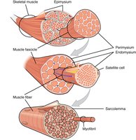
These layers provide support, protection, and pathways for nerves and blood vessels.
Muscle Fiber Structure
A muscle fiber (muscle cell) is a long, cylindrical cell containing multiple nuclei. The plasma membrane is called the sarcolemma, and the cytoplasm is known as sarcoplasm. Muscle fibers contain many myofibrils, which are responsible for the striated appearance and contractile function of the cell.
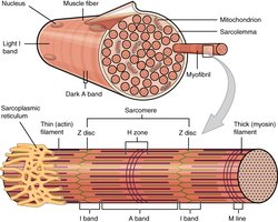
The Sarcomere: Functional Unit of Muscle
Sarcomere Structure
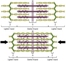
The sarcomere is the basic contractile unit of skeletal muscle, defined as the region between two Z-lines. It contains overlapping thick (myosin) and thin (actin) filaments, which slide past each other during contraction.
Z-line: Defines the boundary of each sarcomere.
I band: Contains only thin filaments.
A band: Contains the entire length of thick filaments.
H zone: Central region with only thick filaments.
M line: Center of the sarcomere.
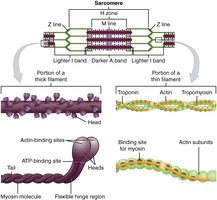
Example: The precise arrangement of actin and myosin filaments allows for efficient muscle contraction and force generation.
Neuromuscular Junction and Muscle Activation
Motor End-Plate and Innervation
The neuromuscular junction (NMJ) is the site where a motor neuron communicates with a muscle fiber. The axon terminal releases the neurotransmitter acetylcholine (ACh), which binds to receptors on the muscle fiber's sarcolemma, initiating an action potential that leads to contraction.
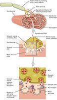
Key Point: The NMJ ensures precise control of muscle contraction by the nervous system.
T-tubules and Sarcoplasmic Reticulum
T-tubules are invaginations of the sarcolemma that conduct electrical impulses deep into the muscle fiber. The sarcoplasmic reticulum (SR) stores calcium ions (Ca2+), which are essential for muscle contraction. The triad consists of a T-tubule flanked by two terminal cisternae of the SR.
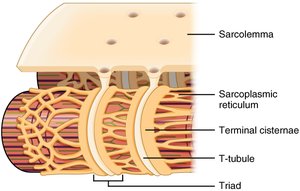
Mechanism of Muscle Contraction
Excitation-Contraction Coupling
When an action potential reaches the NMJ, ACh is released, leading to depolarization of the sarcolemma. This triggers the release of Ca2+ from the SR, which binds to troponin, causing tropomyosin to move and expose binding sites on actin for myosin heads to attach.
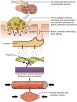
Muscle Relaxation
Relaxation occurs when Ca2+ is pumped back into the SR, causing tropomyosin to cover the actin binding sites. ATP is required for both contraction and relaxation. Muscle fatigue can occur if ATP is depleted.
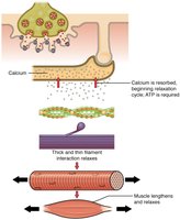
The Sliding Filament Model
During contraction, thin filaments slide past thick filaments, shortening the sarcomere. The Z-lines move closer together, the I band narrows, and the A band remains unchanged.
Cross-Bridge Cycle
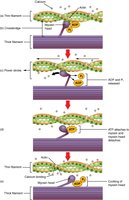
The cross-bridge cycle describes the interaction between actin and myosin:
Calcium binds to troponin, exposing actin binding sites.
Myosin head binds to actin, forming a cross-bridge.
Power stroke: Myosin head pivots, pulling actin filament.
ATP binds to myosin, causing detachment from actin.
ATP hydrolysis re-cocks the myosin head.
Muscle Metabolism
ATP Sources for Muscle Contraction
Muscle fibers require ATP for contraction and relaxation. ATP is generated by:
Creatine Phosphate: Provides a rapid source of ATP for short bursts (about 15 seconds).
Anaerobic Glycolysis: Produces ATP and lactic acid from glucose without oxygen; supports short, intense activity.
Aerobic Respiration: Uses oxygen to produce ATP from glucose, fatty acids, or amino acids; supports prolonged activity.
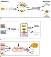
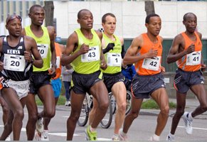
Example: During a sprint, muscles rely on creatine phosphate and anaerobic glycolysis; during a marathon, aerobic respiration predominates.
Types of Muscle Contractions
Isotonic and Isometric Contractions
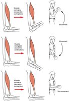
Muscle contractions can be classified as:
Isotonic Contraction: Muscle changes length to move a load (includes concentric and eccentric contractions).
Isometric Contraction: Muscle length does not change; tension increases but the load is not moved.
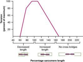
Length-Tension Relationship
Maximum tension is produced when sarcomeres are at an optimal length (80–120% of resting length). Too much or too little overlap between filaments reduces force generation.
Muscle Twitch and Summation
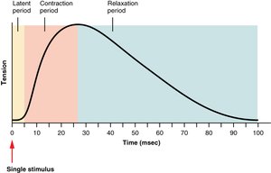
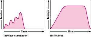
A muscle twitch is a single contraction in response to a single stimulus, consisting of latent, contraction, and relaxation periods. Repeated stimuli can lead to wave summation (increased tension) and tetanus (sustained contraction).
Treppe
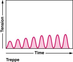
Treppe is the phenomenon where muscle tension increases in a stepwise fashion with repeated stimulation, resembling a staircase pattern.
Muscle Fiber Types and Adaptations
Muscle Fiber Types
There are three main types of skeletal muscle fibers:
Slow Oxidative (SO) Fibers: Fatigue-resistant, use aerobic metabolism, suited for endurance activities.
Fast Oxidative (FO) Fibers: Intermediate properties, use both aerobic and anaerobic metabolism.
Fast Glycolytic (FG) Fibers: Fatigue quickly, use anaerobic metabolism, suited for short, powerful movements.
Muscle Hypertrophy and Atrophy
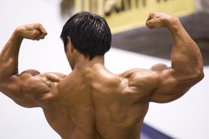
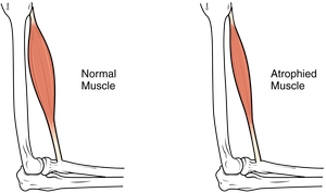
Hypertrophy is the increase in muscle size due to resistance training, primarily by increasing the size of individual muscle fibers. Atrophy is the decrease in muscle mass due to disuse or disease.
Cardiac and Smooth Muscle Tissue
Cardiac Muscle Tissue
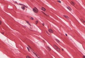
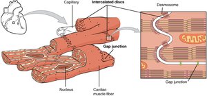
Cardiac muscle is found only in the heart. It is striated, branched, and connected by intercalated discs containing gap junctions and desmosomes, which allow for synchronized contraction.
Smooth Muscle Tissue
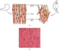
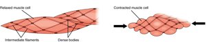
Smooth muscle is found in the walls of hollow organs and is responsible for involuntary movements such as peristalsis. It lacks striations and contracts via a network of dense bodies and intermediate filaments.
Motor Units in Smooth Muscle
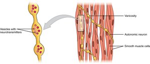
In smooth muscle, autonomic neurons form motor units through structures called varicosities, which release neurotransmitters to stimulate muscle contraction.
Additional info: Cardiac muscle's unique structure allows for rhythmic, coordinated contractions essential for effective heart function, while smooth muscle's arrangement enables sustained contractions in organs without fatigue.
