 뒤로
Muscle Tissue: Structure, Function, and Physiology
스터디 가이드 - 스마트 노트
 자료에 맞춘 맞춤형 노트, 핵심 정의, 예시, 맥락을 확장해 제공합니다.
Muscle Tissue: Structure, Function, and Physiology
Overview of Muscle Types, Special Characteristics, and Functions
Muscles are specialized tissues that convert the energy of ATP into force, enabling movement and stability in the human body. There are three main types of muscle tissue: skeletal, smooth, and cardiac, each with unique structural and functional properties. - Skeletal muscle: Voluntary, striated, attached to bones, responsible for body movement. - Smooth muscle: Involuntary, non-striated, found in walls of hollow organs (e.g., intestines, blood vessels). - Cardiac muscle: Involuntary, striated, found only in the heart. 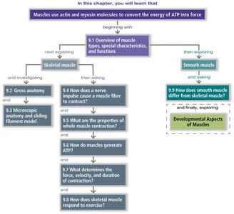
Skeletal Muscle: Gross Anatomy and Microscopic Structure
Skeletal muscle fibers are long, cylindrical cells containing multiple nuclei and are organized into bundles. The microscopic anatomy reveals the presence of myofibrils, sarcomeres, and specialized membranes. - Sarcolemma: The plasma membrane of a muscle fiber. - Myofibrils: Rod-like units within muscle fibers, composed of repeating sarcomeres. - Sarcomere: The functional contractile unit, defined by Z discs and containing thick (myosin) and thin (actin) filaments. 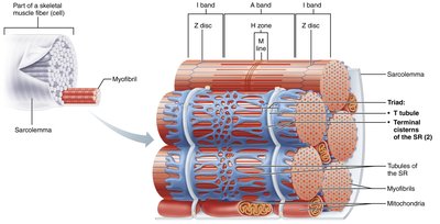
Thin Filament Structure
Thin filaments are primarily composed of actin, along with regulatory proteins tropomyosin and troponin. These proteins control the exposure of myosin-binding sites, regulating muscle contraction. - Actin: Globular protein forming a helical strand. - Tropomyosin: Covers myosin-binding sites on actin in resting muscle. - Troponin: Binds calcium ions, causing tropomyosin to move and expose binding sites. 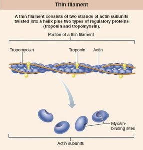
Sarcomere Contraction
Muscle contraction occurs when sarcomeres shorten, as thick and thin filaments slide past each other. - Relaxed sarcomere: Filaments are at maximum separation. - Contracted sarcomere: Filaments overlap, shortening the sarcomere. 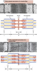
Neuromuscular Junction and Muscle Fiber Activation
The neuromuscular junction is the site where a motor neuron communicates with a muscle fiber, triggering contraction. - Acetylcholine (ACh): Neurotransmitter released from the neuron, binds to receptors on the sarcolemma. - Action potential: Electrical signal generated in the muscle fiber, leading to contraction. 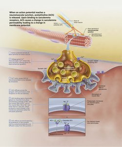
Sequence of Events Leading to Muscle Contraction
Muscle contraction is initiated by a series of steps involving neural stimulation, neurotransmitter release, and calcium ion influx. - Motor neuron activation: Action potential travels down the neuron. - ACh release: Neurotransmitter crosses the synaptic cleft. - Depolarization: Sarcolemma changes membrane potential. - Calcium release: Ca2+ ions released from sarcoplasmic reticulum. - Contraction: Myosin heads bind to actin, pulling filaments. 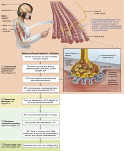
Action Potential and Muscle Fiber Excitation
The action potential is a rapid change in membrane potential, essential for muscle fiber activation. - Depolarization: Voltage-gated Na+ channels open, Na+ enters cell. - Repolarization: Voltage-gated K+ channels open, K+ exits cell. - Resting membrane potential: Maintained by ion gradients. 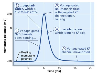
Cross Bridge Cycle: Mechanism of Muscle Contraction
The cross bridge cycle describes the interaction between myosin heads and actin filaments, resulting in muscle contraction. - Attachment: Myosin head binds to actin. - Power stroke: Myosin head pivots, pulling actin. - Detachment: ATP binds to myosin, causing release from actin. - Reactivation: Myosin head resets, ready for another cycle. 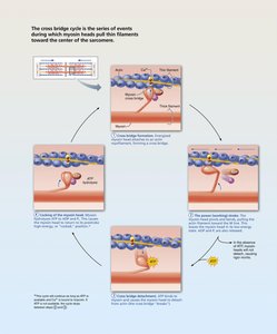
Motor Units and Muscle Fiber Recruitment
A motor unit consists of a motor neuron and all the muscle fibers it innervates. Recruitment of motor units allows for graded muscle responses. - Small motor units: Fine control, fewer fibers per neuron. - Large motor units: Powerful contractions, many fibers per neuron. 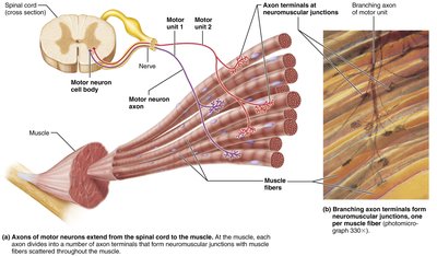
Muscle Twitch and Phases of Contraction
A muscle twitch is the response of a muscle to a single stimulus, consisting of three phases: latent period, contraction, and relaxation. - Latent period: Time between stimulus and contraction onset. - Period of contraction: Muscle tension increases. - Period of relaxation: Muscle tension decreases as Ca2+ is reabsorbed. 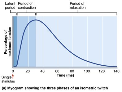
Key Equations
- Force generation: where is the number of active cross bridges, is the force per cross bridge. - Membrane potential: (Nernst equation for potassium ions)
Summary Table: Muscle Fiber Properties
Property | Skeletal Muscle | Smooth Muscle | Cardiac Muscle |
|---|---|---|---|
Control | Voluntary | Involuntary | Involuntary |
Striations | Present | Absent | Present |
Location | Attached to bones | Walls of organs | Heart |
Contraction Speed | Fast | Slow | Intermediate |
Additional info: Academic context was added to clarify the structure and function of muscle tissue, the mechanism of contraction, and the physiological basis of muscle activation.
