 뒤로
Muscle Tissue: Structure, Function, and Physiology
스터디 가이드 - 스마트 노트
 자료에 맞춘 맞춤형 노트, 핵심 정의, 예시, 맥락을 확장해 제공합니다.
Muscle Tissue
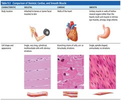
Types of Muscle Tissue
Muscle tissue is classified into three main types: skeletal, cardiac, and smooth. Each type has distinct structural and functional characteristics.
Skeletal Muscle: Attached to bones or skin, responsible for voluntary movements.
Cardiac Muscle: Found only in the heart, responsible for pumping blood.
Smooth Muscle: Located in walls of hollow organs (except the heart), responsible for involuntary movements.
Key Prefixes: Myo-, Mys-, Sarco- are commonly used in muscle terminology (e.g., sarcoplasm, myocyte).
Characteristics & Functions of Muscle Tissue
Muscle tissue exhibits several unique properties and serves essential functions in the body.
Excitability: Ability to receive and respond to stimuli.
Contractility: Ability to shorten forcibly.
Extensibility: Ability to stretch.
Elasticity: Ability to recoil after stretching.
Functions:
Movement
Posture and position
Stability
Thermogenesis (heat production)
Skeletal Muscle Anatomy
Structure and Organization
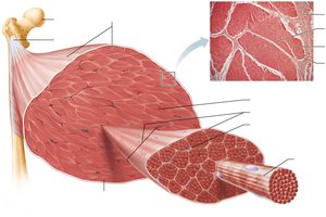
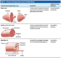
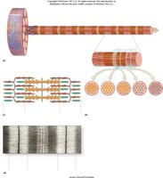
Skeletal muscle is organized into hierarchical structures, each surrounded by connective tissue sheaths.
Epimysium: Surrounds the entire muscle.
Perimysium: Surrounds fascicles (bundles of muscle fibers).
Endomysium: Surrounds individual muscle fibers.
Attachments
Muscles attach to bones via tendons or aponeuroses. The origin is the fixed attachment, while the insertion moves during contraction.
Microanatomy of Skeletal Muscle
Muscle Fiber Structure
Each muscle fiber is a multinucleated cell containing specialized organelles and structures.
Sarcolemma: Plasma membrane of the muscle cell.
Sarcoplasm: Cytoplasm of the muscle cell, rich in glycosomes and myoglobin.
Myofibrils: Rod-like structures responsible for contraction.
Sarcoplasmic Reticulum (SR): Stores and releases calcium ions.
T-Tubules: Invaginations of the sarcolemma that help transmit action potentials.
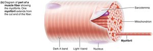
Striations and Bands
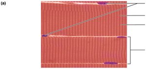
Striations are alternating dark (A band) and light (I band) regions seen in skeletal muscle fibers, resulting from the arrangement of myofilaments.
I band: Isotropic, light band.
A band: Anisotropic, dark band.
Sarcomere Structure
Organization of Myofilaments
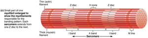
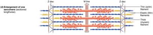
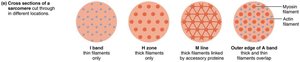
The sarcomere is the functional unit of muscle contraction, defined by the region between two Z-disks.
Actin (thin filament): Anchored to Z-disk.
Myosin (thick filament): Anchored to M-line.
Titin: Elastic filament contributing to extensibility and recoil.
H-zone: Region with only thick filaments.
M-line: Center of the sarcomere.
Molecular Composition of Myofilaments
Myofilaments are composed of contractile and regulatory proteins.
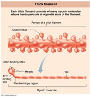
Thick Filament: Made of myosin molecules with heads that bind actin and ATP.
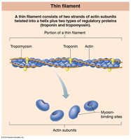
Thin Filament: Made of actin, troponin, and tropomyosin.
Troponin Subunits:
TnI: Inserts on actin.
TnT: Ties to tropomyosin.
TnC: Binds calcium.
Sarcoplasmic Reticulum and T-Tubules
Calcium Storage and Release
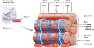
The sarcoplasmic reticulum (SR) stores calcium ions and releases them during muscle contraction. T-tubules help transmit action potentials deep into the muscle fiber.
Terminal cisternae: Enlarged regions of SR adjacent to T-tubules.
Triad: Structure formed by a T-tubule flanked by two terminal cisternae.
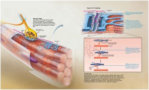
Sliding Filament Model of Contraction
Mechanism of Muscle Contraction
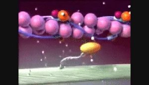
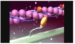
Muscle contraction occurs when myosin heads bind to actin and pull the thin filaments toward the center of the sarcomere, shortening the muscle fiber.
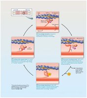
Cross Bridge Formation: Myosin head binds to actin.
Power Stroke: Myosin head pivots, pulling actin.
Detachment: ATP binds to myosin, causing it to release actin.
Cocking: ATP hydrolysis re-energizes myosin head.
Muscle Contraction: The Big Picture
Steps in Skeletal Muscle Contraction
Four main steps are required for skeletal muscle contraction:
Events at the neuromuscular junction
Muscle fiber excitation
Excitation-contraction coupling
Cross bridge cycling
Neuromuscular Junction (NMJ)
The NMJ is the site where a motor neuron communicates with a muscle fiber. Acetylcholine (ACh) is released, binds to receptors, and initiates an action potential in the muscle fiber.
Action potential arrives at axon terminal.
Ca2+ enters, triggering ACh release.
ACh binds to receptors, opening Na+ channels.
End plate potential is generated.
Acetylcholinesterase degrades ACh.
Excitation-Contraction Coupling
Excitation-contraction coupling links the action potential to muscle contraction via calcium release from the SR.
Action potential travels along sarcolemma and T-tubules.
Ca2+ is released from SR.
Ca2+ binds to troponin, shifting tropomyosin and exposing myosin-binding sites.
Cross bridge cycle begins.
Energy for Muscle Contraction
ATP and Muscle Contraction
ATP is essential for muscle contraction, powering cross bridge cycling, calcium pumping, and restoring ionic gradients.
ATP is depleted in 4-6 seconds and must be regenerated.
Three mechanisms for ATP regeneration:
Direct phosphorylation by creatine phosphate (CP)
Anaerobic glycolysis (produces lactic acid)
Aerobic respiration (produces CO2, H2O, and ATP)
Equation for Aerobic Respiration:
Muscle Contraction Types and Responses
Types of Contraction
Muscle contractions can be classified as isometric (no change in length) or isotonic (change in length).
Isotonic: Concentric (shortening) and eccentric (lengthening).
Isometric: Tension increases, but muscle does not shorten.
Motor Units and Muscle Twitch
A motor unit consists of a motor neuron and all the muscle fibers it innervates. A muscle twitch is a single contraction event with three phases: latent, contraction, and relaxation.
Latent period: EC coupling, no tension.
Contraction period: Cross bridge formation, tension increases.
Relaxation period: Ca2+ reenters SR, tension drops.
Graded Muscle Responses
Muscle responses can be graded by changing the frequency or strength of stimulation.
Temporal Summation: Increased frequency leads to tetanus.
Recruitment: Increased strength activates more motor units.
Clinical Considerations
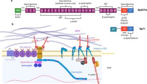
Duchenne Muscular Dystrophy (DMD)
DMD is a genetic disorder caused by a defective dystrophin gene, leading to progressive muscle weakness.
Rigor Mortis
Rigor mortis occurs postmortem due to increased intracellular calcium and lack of ATP, resulting in sustained muscle contraction.
Disuse Atrophy
Muscles must be active to remain healthy. Disuse atrophy results from immobilization or loss of neural stimulation, leading to muscle degeneration.
Problems to Ponder
Why does the A band size remain the same during sarcomere shortening?
What step of the crossbridge cycle is triggered when inorganic phosphate detaches from the myosin head?
What would happen if the SR does not reuptake Ca2+?
Medical Problems to Ponder
How does dantrolene inhibit skeletal muscle contraction and affect body temperature?
How does botulinum toxin affect the neuromuscular junction?
What symptoms might be seen in a patient exposed to curare?
Review: Sequence of Events in Muscle Contraction
Motor neuron action potential
Neurotransmitter release
Muscle cell action potential
Ca2+ release from SR
ATP-powered power stroke
Sliding of myofilaments
Additional info: This study guide covers the essential structure, function, and physiology of muscle tissue, focusing on skeletal muscle. It includes clinical considerations and review questions for exam preparation.
