 뒤로
Ch 9A Muscles and Muscle Tissue: Structure and Function
스터디 가이드 - 스마트 노트
 자료에 맞춘 맞춤형 노트, 핵심 정의, 예시, 맥락을 확장해 제공합니다.
Muscle Tissue Overview
Types of Muscle Tissue
Muscle tissue is essential for movement and comprises nearly half of the body's mass. It transforms chemical energy (ATP) into mechanical energy, enabling force generation. There are three primary types of muscle tissue, each with distinct characteristics and functions.
Skeletal Muscle: Attached to bones or skin, responsible for voluntary movements. Long, cylindrical, striated fibers. Rapid contraction, easily fatigued, powerful.
Cardiac Muscle: Found only in the heart, responsible for pumping blood. Branching, striated cells. Involuntary, contracts at a steady rate.
Smooth Muscle: Located in walls of hollow organs (e.g., stomach, bladder, airways). Single, fusiform, non-striated cells. Involuntary, slow and sustained contractions.
Key Terminology: Prefixes such as myo-, mys-, and sarco- refer to muscle (e.g., sarcoplasm is muscle cell cytoplasm).
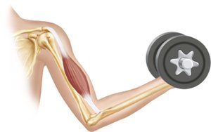
Comparison Table: Skeletal, Cardiac, and Smooth Muscle
Characteristic | Skeletal | Cardiac | Smooth |
|---|---|---|---|
Body Location | Attached to bones or skin | Walls of the heart | Walls of hollow organs |
Cell Shape & Appearance | Long, cylindrical, striated | Branching, striated | Fusiform, non-striated |
Control | Voluntary | Involuntary | Involuntary |
Contraction | Rapid, powerful | Steady, rhythmic | Slow, sustained |
Characteristics and Functions of Muscle Tissue
Four Main Characteristics
All muscle tissues share four fundamental properties:
Excitability (Responsiveness): Ability to receive and respond to stimuli.
Contractility: Ability to shorten forcibly when stimulated.
Extensibility: Ability to be stretched.
Elasticity: Ability to recoil to resting length after stretching.
Four Important Functions
Produce Movement: Responsible for locomotion and manipulation (e.g., walking, digesting, pumping blood).
Maintain Posture and Body Position: Stabilizes the body during activity.
Stabilize Joints: Reinforces and supports joints.
Generate Heat: Muscle contraction produces heat, helping maintain body temperature.
Skeletal Muscle Structure
Gross Structure of Skeletal Muscle
Skeletal muscle is an organ composed of muscle fibers, nerves, blood vessels, and connective tissues. Each muscle receives a nerve, artery, and veins, ensuring adequate oxygen, nutrients, and waste removal.
Connective Tissue Sheaths
Connective tissue sheaths support and reinforce muscle structure. They are organized from external to internal:
Epimysium: Dense irregular connective tissue surrounding the entire muscle; may blend with fascia.
Perimysium: Fibrous connective tissue surrounding fascicles (groups of muscle fibers).
Endomysium: Fine areolar connective tissue surrounding each muscle fiber.
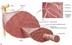
Muscle Attachments
Direct (Fleshy) Attachments: Epimysium fused to periosteum of bone or perichondrium of cartilage.
Indirect Attachments: Connective tissue wrappings extend beyond muscle, forming tendons (ropelike) or aponeuroses (sheetlike).
Microscopic Structure of Skeletal Muscle Fibers
Muscle Fiber Anatomy
Skeletal muscle fibers are long, cylindrical cells with multiple nuclei. The sarcolemma is the plasma membrane, and the sarcoplasm is the cytoplasm, containing glycosomes (for glycogen storage) and myoglobin (for O2 storage).
Specialized Structures
Myofibrils: Densely packed, rodlike elements accounting for ~80% of muscle cell volume. Responsible for striations and contraction.
Sarcoplasmic Reticulum (SR): Network of smooth endoplasmic reticulum tubules surrounding each myofibril. Regulates intracellular Ca2+ levels, stores and releases Ca2+.
T Tubules: Tubular extensions of the sarcolemma that penetrate the cell's interior, allowing electrical signals to reach deep into the muscle fiber.
Myofibril Features
Striations: Alternating dark (A bands) and light (I bands) regions.
Sarcomeres: Smallest contractile units, aligned end-to-end along myofibrils.
Myofilaments: Actin (thin) and myosin (thick) filaments arranged in a specific pattern within sarcomeres.
Molecular Composition of Myofilaments
Thick Filaments: Composed of myosin, with heads forming cross-bridges during contraction.
Thin Filaments: Composed of actin, with regulatory proteins tropomyosin and troponin.
Clinical Application: Duchenne Muscular Dystrophy (DMD)
Overview
DMD is a severe, sex-linked recessive muscular dystrophy affecting mostly males. It is caused by a defective gene for dystrophin, a protein that stabilizes the sarcolemma. The disease leads to muscle degeneration, loss of regenerative capacity, and progressive weakness.
Symptoms: Clumsiness, frequent falls, muscle weakness progressing from extremities to head and chest.
Treatment: No cure; corticosteroids provide temporary relief. Supportive care and recent gene therapy may extend lifespan.
Sarcoplasmic Reticulum and T Tubules
Structure and Function
Sarcoplasmic Reticulum: Surrounds myofibrils, stores and releases Ca2+ for muscle contraction.
T Tubules: Allow electrical impulses to penetrate the muscle fiber, triggering Ca2+ release.
Triad: Consists of a T tubule and two terminal cisterns of the SR, crucial for excitation-contraction coupling.
Summary Table: Structure and Organizational Levels of Skeletal Muscle
Level | Description |
|---|---|
Muscle | Organ composed of bundles of fascicles |
Fascicle | Bundle of muscle fibers |
Muscle Fiber | Single muscle cell |
Myofibril | Rodlike unit within muscle fiber |
Sarcomere | Contractile unit within myofibril |
Myofilament | Actin (thin) and myosin (thick) filaments |
Example: During exercise, skeletal muscle fibers contract rapidly, generating force and heat, while the sarcoplasmic reticulum releases Ca2+ to initiate contraction.
Additional info: The sliding filament model of muscle contraction describes how actin and myosin filaments slide past each other, shortening the sarcomere and producing muscle contraction. The process is regulated by Ca2+ and ATP.
