 뒤로
Muscles and Muscle Tissue: Structure, Function, and Physiology
스터디 가이드 - 스마트 노트
 자료에 맞춘 맞춤형 노트, 핵심 정의, 예시, 맥락을 확장해 제공합니다.
Overview of Muscle Tissue
Types and Functions of Muscle Tissue
Muscle tissue is essential for movement, posture, and various bodily functions. Nearly half of the body's mass is muscle, which transforms chemical energy (ATP) into mechanical energy. There are three main types of muscle tissue, each with distinct characteristics and functions:
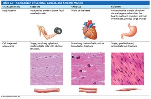
Skeletal Muscle: Attached to bones and skin, responsible for voluntary movements.
Cardiac Muscle: Found only in the heart, responsible for pumping blood.
Smooth Muscle: Located in walls of hollow organs, responsible for involuntary movements.
Muscle tissue shares four main characteristics: excitability, contractility, extensibility, and elasticity. These properties enable muscles to respond to stimuli, contract, stretch, and return to their original shape.
Muscle Terminology
Myo, mys, sarco: Prefixes related to muscle (e.g., sarcoplasm is muscle cell cytoplasm).
Types of Muscle Tissue
Skeletal Muscle
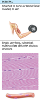
Skeletal muscle tissue is packaged into organs attached to bones and skin. It consists of long, striated fibers and is under voluntary control. Skeletal muscles contract rapidly, tire easily, and are powerful.
Cardiac Muscle
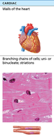
Cardiac muscle tissue is found only in the heart. It is striated and involuntary, contracting at a steady rate due to the heart's pacemaker, but can be influenced by the nervous system.
Smooth Muscle
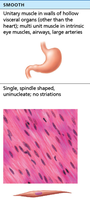
Smooth muscle tissue is located in the walls of hollow organs such as the stomach, urinary bladder, and airways. It is non-striated and involuntary.
Characteristics and Functions of Muscle Tissue
Muscle Characteristics
Excitability: Ability to receive and respond to stimuli.
Contractility: Ability to shorten forcibly when stimulated.
Extensibility: Ability to be stretched.
Elasticity: Ability to recoil to resting length.
Muscle Functions
Produce movement: Responsible for locomotion and manipulation.
Maintain posture: Keeps body position stable.
Stabilize joints: Supports joint integrity.
Generate heat: Maintains body temperature during contraction.
Skeletal Muscle Anatomy
Structure and Organization
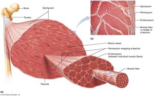
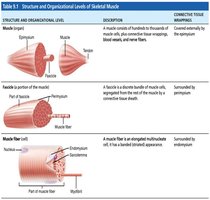
Skeletal muscle is an organ composed of muscle fibers, connective tissue, nerves, and blood vessels. Each muscle receives a nerve, artery, and veins, ensuring proper function and nutrient supply.
Connective Tissue Sheaths
Epimysium: Surrounds entire muscle.
Perimysium: Surrounds fascicles (groups of muscle fibers).
Endomysium: Surrounds individual muscle fibers.
Muscle Attachments
Origin: Attachment to immovable bone.
Insertion: Attachment to movable bone.
Direct attachment: Epimysium fused to periosteum.
Indirect attachment: Connective tissue extends as tendon or aponeurosis.
Muscle Fiber Microanatomy and Sliding Filament Model
Muscle Fiber Structure
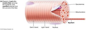
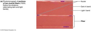
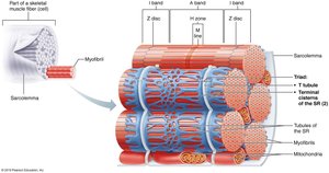
Skeletal muscle fibers are long, cylindrical cells with multiple nuclei. The sarcolemma is the plasma membrane, and the sarcoplasm is the cytoplasm containing glycosomes and myoglobin.
Myofibril Features
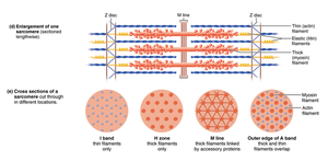
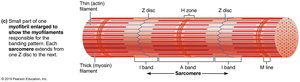
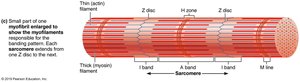
Striations: Alternating dark (A bands) and light (I bands) regions.
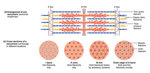
Sarcomeres: Smallest contractile units, aligned end-to-end.
Myofilaments: Actin (thin) and myosin (thick) filaments arranged in a hexagonal pattern.
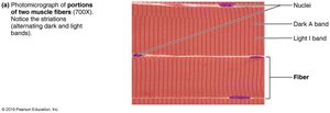
Molecular Composition of Myofilaments
Thick filaments: Composed of myosin molecules with heads that form cross bridges during contraction.
Thin filaments: Composed of actin, tropomyosin, and troponin. Actin has binding sites for myosin heads.
Elastic filament: Titin protein maintains structure and elasticity.
Dystrophin: Links thin filaments to sarcolemma proteins.
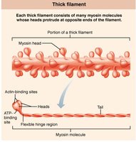
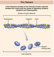
Modified Organelles
Sarcoplasmic Reticulum (SR): Stores and releases Ca2+ for contraction.
T Tubules: Extensions of sarcolemma that transmit electrical signals deep into the fiber.
Triad: Structure formed by a T tubule and two terminal cisterns of SR.
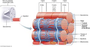
Triad Relationships
T tubule and SR proteins interact to release Ca2+ into the cytoplasm when an electrical impulse arrives, initiating contraction.
Sliding Filament Model of Contraction
During contraction, thin filaments slide past thick filaments, increasing overlap. The process is initiated when myosin heads bind to actin, forming cross bridges. This ratcheting action pulls thin filaments toward the center of the sarcomere, causing muscle shortening.
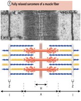
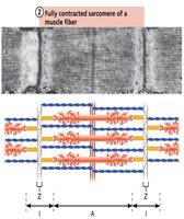
Z discs: Pulled toward M line.
I bands: Shorten.
H zones: Disappear.
A bands: Move closer together.
Muscle Fiber Contraction
Excitable Cells and Action Potentials
Muscle contraction is initiated by signals from motor neurons. Excitable cells can change their membrane potential, and action potentials are transmitted via the neurotransmitter acetylcholine (ACh).
Ion Channels
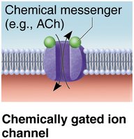
Chemically gated ion channels: Open in response to neurotransmitters (e.g., ACh).
Voltage-gated ion channels: Open in response to changes in membrane voltage.
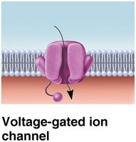
Neuromuscular Junction (NMJ)
The NMJ is the site where motor neurons stimulate muscle fibers. The axon terminal releases ACh into the synaptic cleft, which binds to receptors on the sarcolemma, initiating muscle contraction.
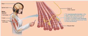
Steps in Muscle Contraction
Events at the neuromuscular junction
Muscle fiber excitation
Excitation-contraction coupling
Cross bridge cycling
Generation of Action Potential Across the Sarcolemma
End plate potential: Local depolarization due to Na+ influx.
Depolarization: If threshold is reached, voltage-gated Na+ channels open, spreading the action potential.
Repolarization: Voltage-gated K+ channels open, restoring resting membrane potential.
Excitation-Contraction (E-C) Coupling
E-C coupling links the action potential to contraction. The AP travels along the sarcolemma and T tubules, triggering Ca2+ release from the SR, which initiates contraction.
Cross Bridge Cycling
Cross bridge formation: Myosin head binds to actin.
Power stroke: Myosin head pivots, pulling actin toward M line.
Detachment: ATP binds to myosin, causing detachment.
Cocking: ATP hydrolysis re-energizes myosin head.
Whole Muscle Contraction
Motor Units
A motor unit consists of a motor neuron and all the muscle fibers it innervates. Smaller motor units allow for finer control.
Muscle Twitch
A muscle twitch is the response of a muscle fiber to a single action potential. It consists of three phases: latent period, contraction, and relaxation.
Graded Muscle Responses
Wave summation: Increased frequency of stimulation increases contraction force.
Recruitment: Increased stimulus strength activates more motor units.
Muscle Tone
Muscle tone is the constant, slightly contracted state of muscles, maintained by spinal reflexes.
Isotonic and Isometric Contractions
Isotonic: Muscle changes length and moves load (concentric or eccentric).
Isometric: Muscle tension increases, but muscle does not change length.
Energy for Contraction and ATP
ATP Regeneration
ATP is essential for muscle contraction and is regenerated by:
Direct phosphorylation: Creatine phosphate donates phosphate to ADP.
Anaerobic pathway: Glycolysis and lactic acid formation.
Aerobic pathway: Cellular respiration in mitochondria.
Muscle Fatigue and Recovery
Fatigue is the inability to contract despite stimulation, caused by ionic imbalances, decreased ATP, and other factors. Recovery requires replenishing oxygen, glycogen, and ATP stores, referred to as excess postexercise oxygen consumption (EPOC).
Tables
Comparison of Skeletal, Cardiac, and Smooth Muscle
Characteristic | Skeletal | Cardiac | Smooth |
|---|---|---|---|
Body Location | Attached to bones or skin | Walls of the heart | Walls of hollow organs |
Cell Shape and Appearance | Long, cylindrical, multinucleate, striated | Branching chains, uni- or binucleate, striated | Spindle-shaped, uninucleate, no striations |
Structure and Organizational Levels of Skeletal Muscle
Structure | Description | Connective Tissue Wrapping |
|---|---|---|
Muscle (organ) | Hundreds to thousands of muscle cells, blood vessels, nerve fibers | Epimysium |
Fascicle | Discrete bundle of muscle cells | Perimysium |
Muscle fiber (cell) | Elongated multinucleate cell | Endomysium |
Key Equations
Direct phosphorylation:
Glycolysis:
Aerobic respiration:
Clinical Context
Duchenne muscular dystrophy: Caused by defective dystrophin gene, leading to muscle degeneration.
Myasthenia gravis: Autoimmune disease affecting ACh receptors, causing muscle weakness.
Rigor mortis: Postmortem muscle stiffness due to lack of ATP and sustained cross bridge formation.
Additional info: Academic context was added to clarify muscle fiber structure, contraction mechanisms, and clinical relevance.
