 뒤로
Muscles Crossing the Hip and Knee Joints: Movements of the Thigh and Leg chapter 10 part 4
스터디 가이드 - 스마트 노트
 자료에 맞춘 맞춤형 노트, 핵심 정의, 예시, 맥락을 확장해 제공합니다.
The Muscular System: Muscles of the Thigh and Leg
Overview of Muscles Crossing the Hip and Knee Joints
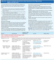
The muscles crossing the hip and knee joints are essential for movements of the thigh and leg, including walking, running, and jumping. These muscles are grouped anatomically and functionally into anterior, medial, and posterior compartments, each with distinct actions and innervations.
Anterior muscles: Flex the femur at the hip and extend the leg at the knee (foreswing of walking).
Posterior muscles: Extend the thigh and flex the leg (backswing of walking).
Medial muscles: Adduct the thigh.
All groups are enclosed by the fascia lata.
Anterior Compartment Muscles
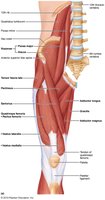
The anterior compartment contains muscles that primarily flex the hip and extend the knee. The quadriceps femoris is the most prominent muscle group in this compartment.
Iliopsoas (iliacus and psoas major): Prime mover of hip flexion.
Tensor fasciae latae: Assists in hip flexion and abduction.
Rectus femoris: Flexes thigh at hip and extends leg at knee.
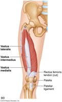
Quadriceps femoris: Consists of four heads (rectus femoris, vastus lateralis, vastus medialis, vastus intermedius); all insert into the quadriceps tendon, patella, and tibial tuberosity. Powerful knee extensor.
Muscle | Description | Origin & Insertion | Action | Nerve Supply |
|---|---|---|---|---|
Rectus femoris | Superficial muscle of anterior thigh | O: Iliac spine; I: Patella/tibial tuberosity | Extends leg, flexes thigh | Femoral nerve |
Vastus lateralis | Lateral thigh muscle | O: Femur; I: Patella/tibial tuberosity | Extends leg, stabilizes knee | Femoral nerve |
Vastus medialis | Medial thigh muscle | O: Femur; I: Patella/tibial tuberosity | Extends leg, stabilizes patella | Femoral nerve |
Vastus intermedius | Deep to rectus femoris | O: Femur; I: Patella/tibial tuberosity | Extends leg | Femoral nerve |
Medial Compartment Muscles
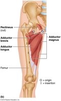
The medial compartment muscles are primarily responsible for adduction of the thigh and also contribute to medial rotation. These muscles are important in movements that press the thighs together.
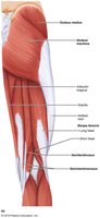
Adductor magnus, longus, brevis: Adduct and medially rotate thigh.
Pectineus: Adducts, flexes, and medially rotates thigh.
Gracilis: Adducts thigh, flexes and medially rotates leg.
Innervated mainly by the obturator nerve.
Muscle | Description | Origin & Insertion | Action | Nerve Supply |
|---|---|---|---|---|
Adductor magnus | Large muscle, medial thigh | O: Ischial/pubic rami; I: Femur | Adducts, flexes, medially rotates thigh | Obturator/sciatic nerves |
Adductor longus | Medial thigh muscle | O: Pubis; I: Femur | Adducts, flexes, medially rotates thigh | Obturator nerve |
Adductor brevis | Short, medial thigh muscle | O: Pubis; I: Femur | Adducts, flexes, medially rotates thigh | Obturator nerve |
Pectineus | Flat, anterior thigh muscle | O: Pubis; I: Femur | Adducts, flexes, medially rotates thigh | Femoral/obturator nerves |
Gracilis | Long, thin, medial thigh muscle | O: Pubis; I: Tibia | Adducts thigh, flexes leg | Obturator nerve |
Posterior Compartment Muscles
The posterior compartment muscles, including the hamstrings, are responsible for extension of the thigh and flexion of the leg. These muscles are crucial for activities such as running and jumping.
Hamstrings: Biceps femoris, semitendinosus, semimembranosus; extend thigh and flex leg.
Innervated by the sciatic nerve.
Common sports injury: "pulled hamstring".
Muscle | Description | Origin & Insertion | Action | Nerve Supply |
|---|---|---|---|---|
Biceps femoris | Lateral hamstring muscle | O: Ischial tuberosity/femur; I: Fibula/tibia | Extends thigh, flexes leg | Sciatic nerve |
Semitendinosus | Medial hamstring muscle | O: Ischial tuberosity; I: Tibia | Extends thigh, flexes leg | Sciatic nerve |
Semimembranosus | Deepest hamstring muscle | O: Ischial tuberosity; I: Tibia | Extends thigh, flexes leg | Sciatic nerve |
Lateral Rotators and Abductors
Lateral rotators and abductors of the thigh stabilize the hip and allow for complex movements such as rotation and abduction. These muscles are important for balance and mobility.
Gluteus maximus: Extends and laterally rotates thigh.
Gluteus medius and minimus: Abduct and medially rotate thigh.
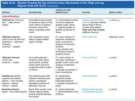
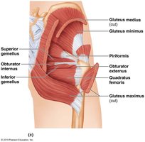
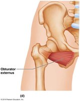
Piriformis, obturator externus/internus, gemellus: Laterally rotate thigh.
Muscle | Description | Origin & Insertion | Action | Nerve Supply |
|---|---|---|---|---|
Gluteus maximus | Largest gluteal muscle | O: Ilium/sacrum/coccyx; I: Femur | Extends, laterally rotates thigh | Inferior gluteal nerve |
Gluteus medius | Middle gluteal muscle | O: Ilium; I: Femur | Abducts, medially rotates thigh | Superior gluteal nerve |
Gluteus minimus | Smallest gluteal muscle | O: Ilium; I: Femur | Abducts, medially rotates thigh | Superior gluteal nerve |
Piriformis | Pyramidal muscle | O: Sacrum; I: Femur | Laterally rotates thigh | Sacral nerves |
Obturator externus/internus | Flat, triangular muscles | O: Obturator foramen; I: Femur | Laterally rotates thigh | Obturator nerve |
Gemellus (superior/inferior) | Small, twin muscles | O: Ischial spine/tuberosity; I: Femur | Laterally rotates thigh | Sacral nerves |
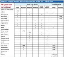
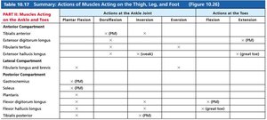
Summary Table: Actions of Muscles Acting on the Thigh, Leg, and Foot
The following table summarizes the actions of major muscles acting on the thigh, leg, and foot, including their roles in flexion, extension, abduction, adduction, rotation, and movements at the knee and ankle joints.
Compartmentalization of Thigh and Leg Muscles
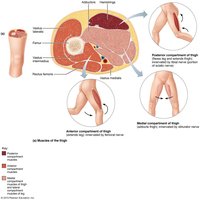
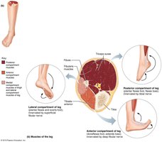
Muscles of the thigh and leg are organized into compartments separated by connective tissue. Each compartment contains muscles with similar functions and innervation.
Anterior compartment of thigh: Extends leg, flexes thigh; innervated by femoral nerve.
Medial compartment of thigh: Adducts thigh; innervated by obturator nerve.
Posterior compartment of thigh: Flexes leg, extends thigh; innervated by sciatic nerve.
Anterior compartment of leg: Dorsiflexes foot; innervated by deep fibular nerve.
Lateral compartment of leg: Everts foot; innervated by superficial fibular nerve.
Posterior compartment of leg: Plantar flexes foot; innervated by tibial nerve.
Key Terms and Concepts
Origin: The fixed attachment point of a muscle.
Insertion: The movable attachment point of a muscle.
Prime mover (agonist): Muscle primarily responsible for a specific movement.
Antagonist: Muscle that opposes the action of the prime mover.
Synergist: Muscle that assists the prime mover.
Fascia lata: Deep fascia enclosing the thigh muscles.
Clinical Relevance
Pulled groin: Injury to overstretched adductor muscles.
Pulled hamstring: Common sports injury involving posterior thigh muscles.
Summary
The muscles crossing the hip and knee joints are essential for locomotion and stability. Understanding their anatomy, actions, and innervation is crucial for comprehending human movement and diagnosing musculoskeletal injuries.
