 뒤로
Muscles Crossing the Shoulder and Elbow Joints: Movements of the Arm and Forearm chapter 10 part 3
스터디 가이드 - 스마트 노트
 자료에 맞춘 맞춤형 노트, 핵심 정의, 예시, 맥락을 확장해 제공합니다.
The Muscular System: Muscles Crossing the Shoulder and Elbow Joints
Overview of Muscles Crossing the Shoulder Joint
The shoulder joint is highly flexible, allowing a wide range of movements of the arm (humerus). Nine muscles cross the shoulder joint, inserting on and moving the humerus. These muscles originate from either the scapula or the axial skeleton and are responsible for actions such as flexion, extension, and adduction. The three prime movers of the arm are the pectoralis major, latissimus dorsi, and deltoid.
Prime Movers: Pectoralis major, latissimus dorsi, deltoid
Rotator Cuff Muscles: Supraspinatus, infraspinatus, teres minor, subscapularis
Synergists: Coracobrachialis, teres major
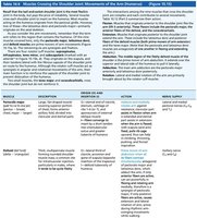
Rotator Cuff Muscles and Their Functions
The rotator cuff muscles act as synergists and fixators, originating on the scapula and reinforcing the shoulder capsule to prevent dislocation. These muscles are essential for stabilizing the shoulder joint during movement.
Supraspinatus: Initiates abduction of the arm; stabilizes shoulder joint
Infraspinatus: Rotates arm laterally
Teres Minor: Rotates arm laterally
Subscapularis: Medially rotates arm
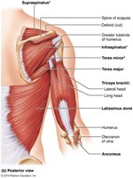
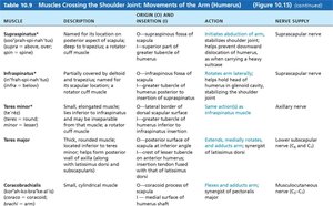
Muscles Crossing the Shoulder Joint: Table Summary
The following table summarizes the muscles crossing the shoulder joint, their descriptions, origins, actions, and nerve supply.
Muscle | Description | Origin (O) and Insertion (I) | Action | Nerve Supply |
|---|---|---|---|---|
Pectoralis major | Large, fan-shaped muscle | O: Clavicle, sternum, ribs; I: Intertubercular sulcus of humerus | Prime mover of arm flexion; adducts, medially rotates arm | Lateral and medial pectoral nerves (C5–C8, T1) |
Latissimus dorsi | Broad, flat, triangular muscle | O: Lower vertebrae, iliac crest, ribs; I: Intertubercular sulcus of humerus | Prime mover of arm extension; adducts, medially rotates arm | Thoracodorsal nerve (C6–C8) |
Deltoid | Thick, multipennate muscle | O: Clavicle, acromion, spine of scapula; I: Deltoid tuberosity of humerus | Prime mover of arm abduction | Axillary nerve (C5, C6) |
Supraspinatus | Named for its location above scapular spine | O: Supraspinous fossa of scapula; I: Greater tubercle of humerus | Initiates abduction of arm | Suprascapular nerve |
Infraspinatus | Partially covered by deltoid and trapezius | O: Infraspinous fossa of scapula; I: Greater tubercle of humerus | Rotates arm laterally | Suprascapular nerve |
Teres minor | Small, elongated muscle | O: Lateral border of scapula; I: Greater tubercle of humerus | Rotates arm laterally | Axillary nerve |
Teres major | Thick, rounded muscle | O: Posterior surface of scapula; I: Intertubercular sulcus of humerus | Extends, medially rotates, and adducts arm | Lower subscapular nerve (C5, C6) |
Coracobrachialis | Small, cylindrical muscle | O: Coracoid process of scapula; I: Medial surface of humerus shaft | Flexes and adducts arm | Musculocutaneous nerve (C5–C7) |
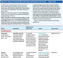
Muscles Crossing the Elbow Joint: Flexion and Extension of the Forearm
The elbow joint is moved by muscles that are divided into anterior and posterior compartments. The anterior muscles are primarily flexors, while the posterior muscles are extensors. The walls of fascia divide the arm into these two compartments.
Anterior Muscles: Biceps brachii, brachialis, brachioradialis
Posterior Muscles: Triceps brachii, anconeus
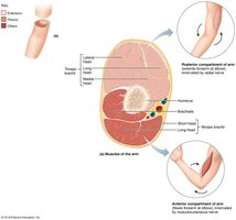
Muscles Crossing the Elbow Joint: Table Summary
The following table summarizes the muscles crossing the elbow joint, their descriptions, origins, actions, and nerve supply.
Muscle | Description | Origin (O) and Insertion (I) | Action | Nerve Supply |
|---|---|---|---|---|
Biceps brachii | Two-headed fusiform muscle | O: Short head—coracoid process; long head—supraglenoid tubercle; I: Radial tuberosity | Flexes and supinates forearm | Musculocutaneous nerve (C5–C6) |
Brachialis | Strong muscle deep to biceps brachii | O: Distal humerus; I: Coronoid process of ulna | Major forearm flexor | Musculocutaneous nerve |
Brachioradialis | Superficial muscle of lateral forearm | O: Lateral supracondylar ridge of humerus; I: Styloid process of radius | Synergist in flexing forearm | Radial nerve |
Triceps brachii | Large fleshy muscle; three heads | O: Long head—infraglenoid tubercle; lateral head—posterior shaft of humerus; medial head—posterior shaft of humerus; I: Olecranon of ulna | Powerful forearm extensor | Radial nerve (C6–C8) |
Anconeus | Short triangular muscle | O: Lateral epicondyle of humerus; I: Olecranon of ulna | Synergist in extension of forearm | Radial nerve |
Muscles of the Forearm: Movements of the Wrist, Hand, and Fingers
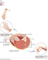
The forearm muscles are divided into anterior and posterior compartments. Most anterior muscles are flexors and insert via the flexor retinaculum, while most posterior muscles are extensors and insert via the extensor retinaculum. These muscles are further divided into superficial and deep groups.
Flexors: Flex wrist, fingers, and thumb
Extensors: Extend wrist, fingers, and thumb
Pronators: Pronator teres and pronator quadratus pronate forearm
Supinators: Supinator acts as a synergist with biceps brachii in forearm supination
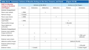
Summary Table: Actions of Muscles Acting on the Arm, Forearm, and Hand
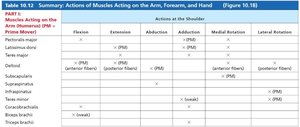
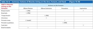
The following tables summarize the actions of muscles acting on the arm, forearm, and hand, including flexion, extension, abduction, adduction, and rotation at various joints.
Muscle | Flexion | Extension | Abduction | Adduction | Medial Rotation | Lateral Rotation |
|---|---|---|---|---|---|---|
Pectoralis major | x (PM) | x (PM) | ||||
Latissimus dorsi | x (PM) | x (PM) | x (PM) | |||
Deltoid | x (anterior fibers) | x (posterior fibers) | x (PM) | x (anterior fibers) | x (posterior fibers) | |
Supraspinatus | x (PM) | |||||
Infraspinatus | x (PM) | |||||
Teres minor | x (PM) | |||||
Subscapularis | x (PM) | |||||
Coracobrachialis | x (weak) | x (weak) | ||||
Triceps brachii |
Muscle | Elbow Flexion | Elbow Extension | Pronation | Supination |
|---|---|---|---|---|
Biceps brachii | x (PM) | x | ||
Brachialis | x (PM) | |||
Brachioradialis | x | |||
Triceps brachii | x (PM) | |||
Anconeus | x | |||
Pronator teres | x (weak) | |||
Pronator quadratus | x (PM) | |||
Supinator | x |
Muscle | Wrist Flexion | Wrist Extension | Abduction | Adduction | Finger Flexion | Finger Extension |
|---|---|---|---|---|---|---|
Flexor carpi radialis | x (PM) | x | ||||
Flexor carpi ulnaris | x (PM) | x | ||||
Palmaris longus | x (PM) | |||||
Flexor digitorum superficialis | x (PM) | x (PM) | ||||
Flexor digitorum profundus | x (PM) | x (PM) | ||||
Extensor carpi radialis longus | x (PM) | x | ||||
Extensor carpi radialis brevis | x (PM) | x | ||||
Extensor carpi ulnaris | x (PM) | x | ||||
Extensor digitorum | x (PM) | x (PM) | ||||
Abductor pollicis longus | x (thumb) | |||||
Extensor pollicis longus | x (thumb) | |||||
Extensor indicis | x (index finger) |
Key Terms and Concepts
Flexion: Decreasing the angle between two bones
Extension: Increasing the angle between two bones
Abduction: Movement away from the midline
Adduction: Movement toward the midline
Medial Rotation: Rotating toward the midline
Lateral Rotation: Rotating away from the midline
Synergist: Muscle that assists a prime mover
Fixator: Muscle that stabilizes the origin of the prime mover
Example: Application in Movement
During a push-up, the pectoralis major and triceps brachii are activated to flex and extend the arm at the shoulder and elbow joints, respectively. The rotator cuff muscles stabilize the shoulder joint, preventing dislocation.

Additional info:
Tables and images were interpreted and grouped logically to provide a comprehensive overview of the muscles acting on the shoulder, elbow, and forearm.
Muscle actions were expanded with academic context for clarity and completeness.
