 뒤로
Muscles of the Body: Structure, Function, and Organization
스터디 가이드 - 스마트 노트
 자료에 맞춘 맞춤형 노트, 핵심 정의, 예시, 맥락을 확장해 제공합니다.
Muscles of the Body: Overview
General Organization of Skeletal Muscles
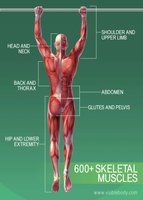
Skeletal muscles are distributed throughout the body and are responsible for voluntary movements, posture, and support. There are over 600 skeletal muscles, each classified by their location and function.
Head and Neck: Muscles involved in facial expression, mastication, and head movement.
Back and Thorax: Muscles supporting the spine and aiding in respiration.
Shoulder and Upper Limb: Muscles responsible for movement of the arm, forearm, and hand.
Abdomen: Muscles involved in trunk movement and protection of internal organs.
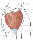
Glutes and Pelvis: Muscles supporting pelvic organs and movement of the thigh.
Hip and Lower Extremity: Muscles for locomotion and stability.
Structural Organization of Skeletal Muscles
Connective Tissue Membranes
Skeletal muscles are organized into layers of connective tissue that provide structure and support:
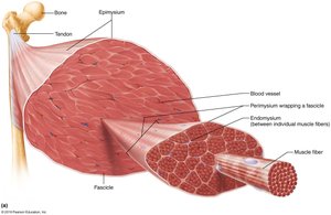
Endomysium: Fine layer of areolar connective tissue surrounding each muscle fiber (cell).
Perimysium: Dense irregular connective tissue wrapping around fascicles (groups of muscle fibers).
Epimysium: Dense, irregular fibrous connective tissue surrounding the whole muscle.
Deep fascia: Coarser layer binding muscles into functional groups (e.g., hamstrings vs. quadriceps).
Each muscle fiber is a single muscle cell, and the connective tissue wrappings help transmit force and protect muscle tissue.
Muscle Attachments
Direct vs. Indirect Attachments
Muscles attach to bones either directly or indirectly:
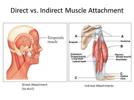
Direct Attachment: Epimysium fused to periosteum of bone or perichondrium of cartilage (e.g., temporalis muscle).
Indirect Attachment: Most common; involves tendons (cordlike structures) or aponeuroses (broad, flat sheets).
Muscles span joints and attach to bones at two main points:
Origin: The stationary attachment point.
Insertion: The movable attachment point.
Functional Groups of Muscles
Agonist, Antagonist, Synergist, and Fixator
Muscles are classified based on their roles in movement:
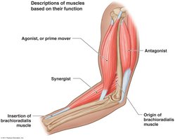
Agonist (Prime Mover): Provides the major force for a particular movement (e.g., brachialis in elbow flexion).
Antagonist: Opposes or reverses a particular movement; provides resistance and control (e.g., triceps brachii in elbow flexion).
Synergist: Assists the prime mover by promoting the same movement or reducing unnecessary movement.
Fixator: A type of synergist that immobilizes a bone, stabilizing the origin of the prime mover.
Muscles can change their role depending on the movement being performed.
Arrangement of Fascicles
Types of Fascicle Arrangements
The arrangement of fascicles within a muscle determines its range of motion and power:
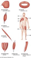
Circular: Fascicles arranged in concentric rings (e.g., orbicularis oculi).
Parallel: Fascicles parallel to the long axis of the muscle (e.g., sartorius).
Fusiform: Spindle-shaped with a central belly (e.g., biceps brachii).
Pennate: Fascicles attach obliquely to a central tendon; can be unipennate, bipennate, or multipennate (e.g., deltoid).
Convergent: Fascicles converge toward a single tendon insertion (e.g., pectoralis major).
Respiratory Muscles
Intercostal Muscles and Diaphragm
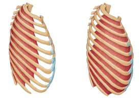
Muscles involved in respiration include the intercostal muscles and the diaphragm:
External Intercostals: Elevate the rib cage, aiding in inspiration.
Internal Intercostals: Depress the rib cage, aiding in forced expiration.
Diaphragm: Dome-shaped muscle; prime mover of inspiration, flattens to increase thoracic volume.
Muscles of Head and Neck
Muscles of Facial Expression
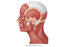
Muscles of facial expression insert into skin or other muscles and are innervated by the facial nerve (CN VII). Examples include:
Epicranius: Main muscle of the scalp, includes frontal and occipital bellies.
Orbicularis oculi: Closes the eye, blinking, squinting.
Zygomaticus: Raises the corners of the mouth (smiling).
Orbicularis oris: Closes, purses, and protrudes lips.
Mentalis: Protrudes lower lip, wrinkles chin.
Buccinator: Holds food in place during chewing.
Platysma: Tenses skin of neck, helps depress mandible.
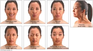
Muscles of Mastication and Tongue Movement
Muscles of Mastication
Four pairs of muscles are involved in mastication, all innervated by the mandibular division of cranial nerve V:
Temporalis: Elevates and retracts mandible.
Masseter: Prime mover of jaw closure.
Medial and Lateral Pterygoids: Side-to-side grinding movements.
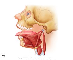
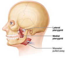
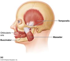
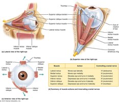
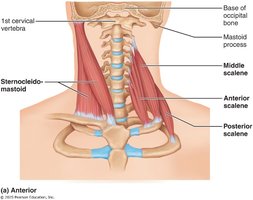
Muscles of the Neck and Vertebral Column
Head and Neck Movements
Muscles originating from the axial skeleton move the head and neck:
Sternocleidomastoid: Prime mover of head flexion; rotates head to opposite side.
Scalenes: Elevate first two ribs, aid in inspiration, flex and rotate neck.
Splenius: Extends or rotates head.
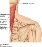
Muscles of the Vertebral Column
These muscles maintain posture and allow movement of the spine:
Erector Spinae: Prime mover of back extension; consists of iliocostalis, longissimus, and spinalis.
Quadratus Lumborum: Flexes vertebral column laterally; extends lumbar spine.
Semispinalis: Extends vertebral column and head; rotation of head.
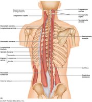
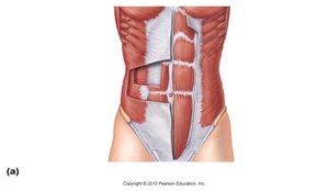
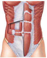
Muscles of the Abdominal Wall
Abdominal Muscles
Four paired muscles form the lateral and anterior abdominal wall:
Internal Oblique
External Oblique
Transversus Abdominis
Rectus Abdominis
These muscles provide trunk movement, support viscera, and aid in actions such as urination, defecation, childbirth, vomiting, coughing, and screaming.

Summary Table: Connective Tissue Layers of Skeletal Muscle
Layer | Description | Function |
|---|---|---|
Endomysium | Surrounds each muscle fiber | Provides support and insulation |
Perimysium | Wraps fascicles (groups of fibers) | Bundles fibers for coordinated contraction |
Epimysium | Surrounds entire muscle | Protects and supports muscle as a whole |
Deep fascia | Binds muscles into groups | Organizes muscles for functional movement |
Key Terms and Principles
Agonist: Main muscle responsible for movement.
Antagonist: Muscle that opposes the agonist.
Synergist: Muscle that assists the agonist.
Fixator: Muscle that stabilizes the origin.
Origin: Stationary attachment point.
Insertion: Movable attachment point.
Formulas and Equations
Muscle force transmission is based on the arrangement of connective tissue and muscle fibers. The total force generated by a muscle is proportional to the number of fibers contracting:
Muscle contraction follows the sliding filament theory:
Additional info: The notes above expand on brief points from the original material, providing definitions, examples, and academic context for each topic. Images included are directly relevant to the anatomical structures and functions discussed.
