 뒤로
Muscles of the Body: Structure, Function, and Key Anatomical Features
스터디 가이드 - 스마트 노트
 자료에 맞춘 맞춤형 노트, 핵심 정의, 예시, 맥락을 확장해 제공합니다.
Muscle Tissue and Muscles of the Body
Overview of Muscle Structure and Function
Muscles are specialized tissues responsible for movement, posture, and various physiological functions. They operate by contracting and relaxing, which enables the body to perform actions such as flexion, extension, abduction, and adduction. Understanding the origin, insertion, and action of each muscle is fundamental in anatomy and physiology.
Origin: The fixed attachment point of the muscle.
Insertion: The movable attachment point where the muscle exerts its action.
Action: The movement produced when the muscle contracts.
Patterns of Fascicle Arrangement in Muscles
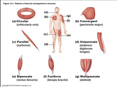
Muscle fascicles are bundles of muscle fibers arranged in specific patterns, which influence the muscle's function and strength. The arrangement determines the range of motion and force generation.
Circular: Fascicles arranged in concentric rings (e.g., orbicularis oris).
Convergent: Fascicles converge toward a single tendon (e.g., pectoralis major).
Parallel: Fascicles run parallel to the long axis (e.g., sartorius).
Unipennate: Fascicles insert into one side of the tendon (e.g., extensor digitorum longus).
Bipennate: Fascicles insert into both sides of the tendon (e.g., rectus femoris).
Fusiform: Spindle-shaped with a central belly (e.g., biceps brachii).
Multipennate: Multiple tendons with fascicles arranged around them (e.g., deltoid).
Major Muscles of the Body
Head and Neck Muscles
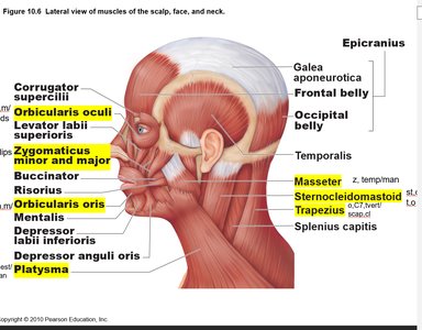
Muscles in the head and neck are essential for facial expression, mastication, and movement of the head.
Orbicularis oculi: Closes the eyelids; origin at frontal and maxilla bones, insertion at eyelids.
Orbicularis oris: Purses lips; origin at maxilla and mandible, insertion at lips.
Zygomaticus major and minor: Elevate corners of mouth for smiling; origin at zygomatic bone, insertion at corners of mouth.
Masseter: Prime mover of jaw closure; origin at zygomatic bone and arch, insertion at mandible.
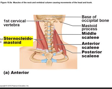
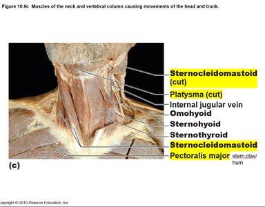
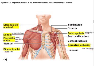
Sternocleidomastoid: Flexes and rotates neck; origin at sternum and clavicle, insertion at mastoid process.
Platysma: Tenses skin of neck and depresses corners of mouth; origin at chest, insertion at mandible.
Thorax Muscles
Muscles of the thorax are involved in movements of the shoulder and arm, as well as respiration.
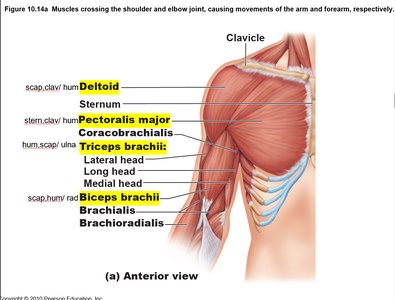
Pectoralis major: Flexes, adducts, and medially rotates the arm; origin at sternum and clavicle, insertion at humerus.
Serratus anterior: Protracts and holds scapula against chest wall; origin at ribs 1-8, insertion at scapula.
Abdominal Muscles
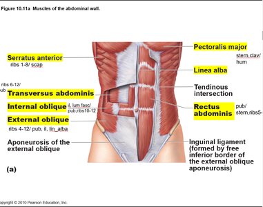
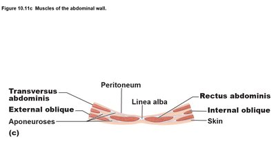
The abdominal muscles support the trunk, allow movement, and help maintain posture. They also protect internal organs and assist in functions such as respiration and defecation.
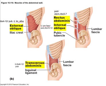
Rectus abdominis: Flexes the spine; origin at pubic bone, insertion at sternum and ribs 5-7.
Transversus abdominis: Compresses abdominal contents; origin at ilium and ribs 6-12, insertion at pubic bone and linea alba.
Internal oblique: Compresses abdominal wall, rotates and laterally flexes abdomen; origin at ilium and lumbar fascia, insertion at pubic bone and ribs 10-12.
External oblique: Compresses abdominal wall, rotates and laterally flexes abdomen; origin at ribs 4-12, insertion at pubic bone, ilium, and linea alba.
Linea alba: Tendinous structure running down the midline of the abdomen, formed by the fusion of aponeuroses.
Shoulder and Arm Muscles
These muscles are responsible for movements of the shoulder and upper limb, including abduction, flexion, extension, and rotation.
Deltoid: Abducts the arm; origin at scapula and clavicle, insertion at humerus.
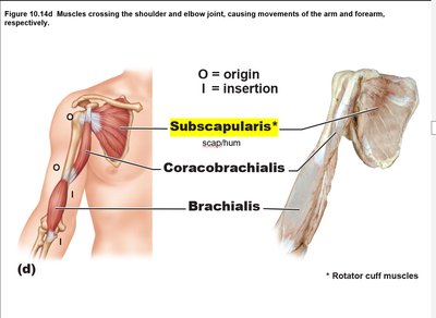
Subscapularis: Medially rotates the humerus; origin at anterior scapula, insertion at humerus.
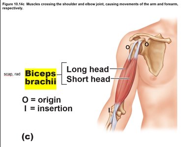
Biceps brachii: Flexes the arm; origin at scapula, insertion at radius.
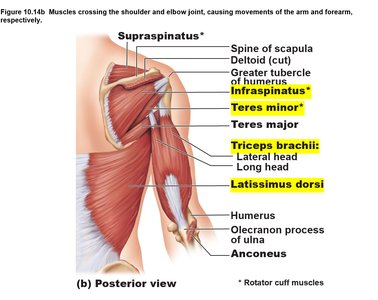
Triceps brachii: Extends the arm; origin at humerus and scapula, insertion at ulna.
Back Muscles
Muscles of the back stabilize and move the scapula and shoulder, and are involved in movements of the upper limb.
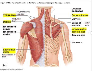
Trapezius: Stabilizes, raises, rotates, and retracts scapula; origin at occipital bone, C7, and thoracic vertebrae, insertion at scapula and clavicle.
Latissimus dorsi: Adducts, extends, and medially rotates the arm; origin at thoracic and lumbar vertebrae, insertion at humerus.
Infraspinatus: Lateral rotation of arm; origin at scapula, insertion at humerus.
Supraspinatus: Initiates abduction of humerus; origin at scapula, insertion at humerus.
Teres minor: Lateral rotation of arm; origin at scapula, insertion at humerus.
Forearm Muscles
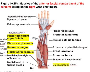
Forearm muscles control movements of the wrist, hand, and fingers. They are classified as flexors (anterior) and extensors (posterior).
Flexor digitorum superficialis: Flexes fingers; origin at humerus, radius, and ulna, insertion at phalanges 2-5.
Flexor carpi ulnaris: Flexes wrist; origin at humerus and ulna, insertion at metacarpal #5, pisiform, and hamate bones.
Palmaris longus: Tenses skin and fascia of palm; origin at humerus, insertion at palmar aponeurosis.
Flexor carpi radialis: Flexes wrist; origin at humerus, insertion at metacarpals 2 and 3.
Extensor carpi radialis longus: Extends and abducts hand; origin at humerus, insertion at metacarpal #2.
Extensor digitorum: Extends fingers; origin at humerus, insertion at phalanges 2-5.
Summary Table: Major Muscles, Origins, Insertions, and Actions
The following table summarizes the key muscles discussed, their origins, insertions, and primary actions.
Muscle | Origin | Insertion | Action |
|---|---|---|---|
Orbicularis oculi | Frontal bone, maxilla | Eye lids | Closes eyes |
Orbicularis oris | Maxilla, mandible | Lips | Purses lips |
Zygomaticus major/minor | Zygomatic bone | Corners of mouth | Smiling |
Masseter | Zygomatic bone/arch | Mandible | Jaw closure |
Sternocleidomastoid | Sternum, clavicle | Mastoid process | Neck flexion/rotation |
Platysma | Chest | Mandible | Tenses neck skin |
Pectoralis major | Sternum, clavicle | Humerus | Arm flexion/adduction |
Serratus anterior | Ribs 1-8 | Scapula | Protracts scapula |
Rectus abdominis | Pubic bone | Sternum, ribs 5-7 | Spine flexion |
Transversus abdominis | Ilium, ribs 6-12 | Pubic bone, linea alba | Compresses abdomen |
Internal oblique | Ilium, lumbar fascia | Pubic bone, ribs 10-12 | Compresses/rotates abdomen |
External oblique | Ribs 4-12 | Pubic bone, ilium, linea alba | Compresses/rotates abdomen |
Deltoid | Scapula, clavicle | Humerus | Abducts arm |
Subscapularis | Scapula | Humerus | Medial rotation |
Biceps brachii | Scapula | Radius | Flexes arm |
Triceps brachii | Humerus, scapula | Ulna | Extends arm |
Trapezius | Occipital bone, C7, thoracic vertebrae | Scapula, clavicle | Stabilizes scapula |
Latissimus dorsi | Thoracic/lumbar vertebrae | Humerus | Adducts/extends arm |
Infraspinatus | Scapula | Humerus | Lateral rotation |
Supraspinatus | Scapula | Humerus | Abduction |
Teres minor | Scapula | Humerus | Lateral rotation |
Flexor digitorum superficialis | Humerus, radius, ulna | Phalanges 2-5 | Flexes fingers |
Flexor carpi ulnaris | Humerus, ulna | Metacarpal #5, pisiform, hamate | Flexes wrist |
Palmaris longus | Humerus | Palmar aponeurosis | Tenses palm |
Flexor carpi radialis | Humerus | Metacarpals 2, 3 | Flexes wrist |
Extensor carpi radialis longus | Humerus | Metacarpal #2 | Extends/abducts hand |
Extensor digitorum | Humerus | Phalanges 2-5 | Extends fingers |
Examples and Applications
Flexion and Extension: Biceps brachii flexes the forearm, while triceps brachii extends it.
Abduction and Adduction: Deltoid abducts the arm, pectoralis major adducts the arm.
Rotation: Subscapularis and infraspinatus rotate the humerus medially and laterally, respectively.
Additional info:
Muscle actions are often described relative to anatomical position.
Synergists assist prime movers, while antagonists oppose them.
Muscle names often reflect their location, shape, or function.
