 뒤로
뒤로Muscles of the Body: Structure, Function, and Organization
스터디 가이드 - 스마트 노트
 자료에 맞춘 맞춤형 노트, 핵심 정의, 예시, 맥락을 확장해 제공합니다.
Mechanics of Muscle Function
Overview of Skeletal Muscle Function
Skeletal muscles attach to the bony skeleton and are responsible for producing movements. The physical parameters that define how a muscle acts on the skeleton include the arrangement of fascicles within a muscle, principles of lever mechanics, and the position of a muscle as it crosses a joint.
Fascicle arrangement determines the action and efficiency of a muscle.
Lever mechanics describe how bones and muscles interact to produce movement.
Muscle position relative to a joint influences the type of movement produced.
Arrangement of Fascicles in Muscles
Types of Fascicle Arrangement
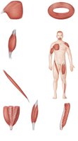
Fascicles are bundles of muscle fibers arranged in specific patterns, which influence muscle action.
Circular: Fascicles arranged in concentric rings, typically surrounding external body openings. These muscles are called sphincters (e.g., orbicularis oris, orbicularis oculi).
Convergent: Broad origin with fascicles converging toward a single tendon (e.g., pectoralis major).
Parallel: Fascicles run parallel to the long axis of the muscle. Includes straplike (e.g., sartorius) and fusiform (e.g., biceps brachii) muscles.
Pennate: Fascicles attach obliquely to a tendon. Types include unipennate (one side), bipennate (both sides), and multipennate (multiple sides, e.g., deltoid).
Functional Implications
Muscles with long, parallel fibers have a greater range of motion.
Pennate muscles contain more fibers and are more powerful.
Lever Systems: Bone-Muscle Relationships
Principles of Leverage
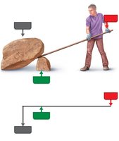
Movement of skeletal muscles involves leverage, which is described by the following components:
Lever: A rigid bar (bone) that moves.
Fulcrum: A fixed point (joint).
Effort: The applied force (muscle contraction).
Load: The resistance (body part or object moved).
Mechanical Advantage and Disadvantage
Mechanical advantage: Effort arm is longer than load arm, allowing a smaller force to move a larger load.
Mechanical disadvantage: Effort arm is shorter than load arm, requiring more force but increasing speed and range of movement.
Classes of Levers
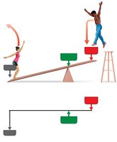
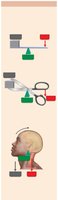
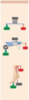
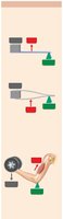
First-class lever: Fulcrum is between effort and load (e.g., seesaw, scissors, lifting head off chest).
Second-class lever: Load is between fulcrum and effort (e.g., wheelbarrow, standing on tip-toe).
Third-class lever: Effort is between fulcrum and load (e.g., tweezers, flexing forearm by biceps brachii).
Muscle Actions and Interactions
Types of Muscle Roles
Muscles work in groups to produce and control movement.
Prime mover (agonist): Main muscle responsible for a movement.
Antagonist: Opposes or reverses the action of the prime mover.
Synergist: Assists the prime mover by adding force or reducing unwanted movements.
Fixator: Stabilizes a bone or joint during movement.
Muscle Position and Movement
Muscles crossing the anterior side of a joint produce flexion.
Muscles crossing the posterior side produce extension.
Lateral crossing produces abduction; medial crossing produces adduction.
Organization Scheme Based on Embryonic Development
Muscle Development and Groups
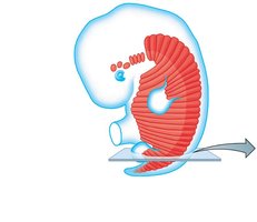
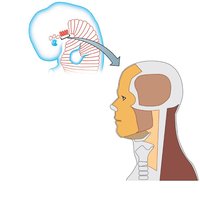
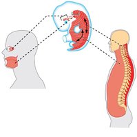
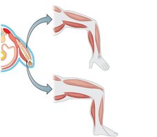
Muscles are organized into developmental and functional groups. They develop from mesodermal structures called myotomes and somitomeres.
Visceral organ muscles: Smooth and cardiac muscle from splanchnic mesoderm.
Pharyngeal arch muscles: Skeletal muscles of the pharynx, head, and neck from 4th–7th somitomeres.
Axial muscles: Muscles of the trunk, neck, and some head muscles from myotomes and somitomeres.
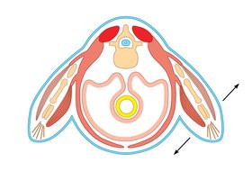
Limb muscles: Arise from lateral parts of myotomes; extensors and flexors are organized based on their position relative to limb bones.
Muscle Compartments of the Limbs
Compartmental Organization
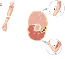
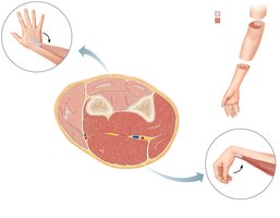
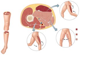
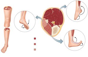
Dense connective tissue divides limb muscles into compartments, each with similar actions and innervation.
Muscles in the same compartment act as synergists.
Opposing compartments contain agonist-antagonist pairs.
Each compartment is innervated by a single nerve.
Upper Limb Compartments
Anterior arm: Flexes shoulder/forearm; innervated by musculocutaneous nerve.
Posterior arm: Extends forearm; innervated by radial nerve.
Anterior forearm: Flexes wrist/digits; innervated by median/ulnar nerve.
Posterior forearm: Extends wrist/fingers; innervated by radial nerve.
Thigh and Leg Compartments
Thigh: Posterior (extends hip/flexes knee, tibial nerve), anterior (flexes hip/extends knee, femoral nerve), medial (adducts thigh, obturator nerve).
Leg: Posterior (plantar flexes/flexes toes, tibial nerve), anterior (dorsiflexes/extends toes, deep fibular nerve), lateral (plantar flexes/everts foot, superficial fibular nerve).
Naming the Skeletal Muscles
Criteria for Muscle Names
Muscles are named based on several characteristics:
Location: e.g., brachialis (arm).
Shape: e.g., deltoid (triangular).
Size: e.g., gluteus maximus, minimus.
Direction of fibers: e.g., rectus abdominis, transversus abdominis.
Attachment points: e.g., brachioradialis.
Number of origins: e.g., biceps, triceps, quadriceps.
Action: e.g., flexor, extensor, adductor, abductor.
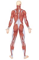
Muscles of the Head—Facial Expression
Facial Muscles
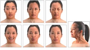
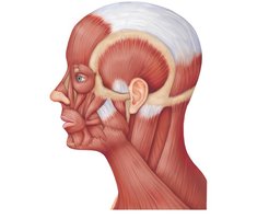
Muscles of facial expression are thin, variable in shape, and often insert into the skin rather than bones. They are innervated by the facial nerve (cranial nerve VII).
Examples include frontal belly of epicranius, corrugator supercilii, orbicularis oculi, zygomaticus major, orbicularis oris, mentalis, and platysma.
Muscles of Mastication and Tongue Movement
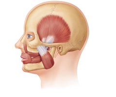
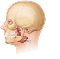
Mastication Muscles
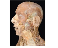
Four main pairs of muscles are involved in mastication, innervated by the mandibular division of the trigeminal nerve (cranial nerve V).
Prime movers of jaw closure: masseter and temporalis.
Side-to-side movement: pterygoid muscles.
Compression of cheeks: buccinator muscles.
Extrinsic Muscles of the Tongue
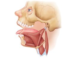
These muscles move the tongue in various directions and are innervated by the hypoglossal nerve (cranial nerve XII).
Include genioglossus, styloglossus, and hyoglossus.
Muscles of the Anterior Neck and Throat—Swallowing
Neck Muscles for Swallowing
The neck is divided into anterior and posterior triangles by the sternocleidomastoid. The anterior triangle contains suprahyoid and infrahyoid muscles, which participate in swallowing. Pharyngeal constrictors squeeze food into the esophagus. 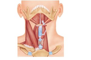
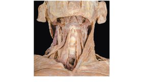
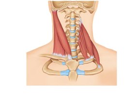
Muscles of the Neck and Vertebral Column
Head and Neck Movement
Flexion: sternocleidomastoid.
Lateral flexion: splenius capitis, splenius cervicis.
Extension: trapezius, splenius.
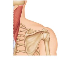
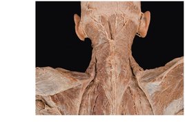
Trunk Extension
Deep muscles of the back, including the erector spinae group, maintain normal curvatures of the spine and form a column from sacrum to skull. 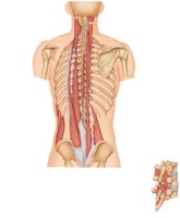
Deep Muscles of the Thorax—Breathing
Intercostal Muscles
Both external and internal intercostals are involved in inspiration and active expiration. Accessory muscles of ventilation are recruited during forced breathing. 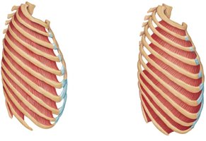
Diaphragm
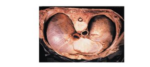
The diaphragm is the most important muscle of respiration, flattening as it contracts to increase thoracic cavity volume. 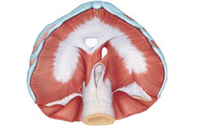
Muscles of the Abdominal Wall
Abdominal Wall Structure
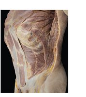
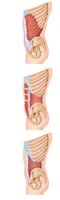
The lateral and anterior abdominal wall is formed from three flat muscle sheets: external oblique, internal oblique, and transversus abdominis. The rectus abdominis inserts at the linea alba. 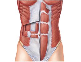

Muscles of the Pelvic Floor and Perineum
Pelvic Diaphragm
The pelvic floor is a sheet of two muscles (levator ani and coccygeus) that support pelvic organs. 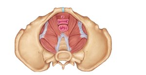
Perineum
The urogenital diaphragm is formed from sphincter urethrae and deep transverse perineus. 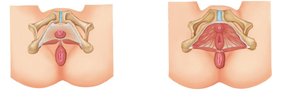
Superficial Perineal Muscles
Includes ischiocavernosus, bulbospongiosus, and superficial transverse perineus. 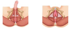
Superficial Muscles of the Thorax
Movements of the Scapula
Muscles such as pectoralis major, pectoralis minor, serratus anterior, and subclavius move the scapula. 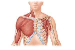
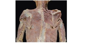
Posterior Thorax Muscles
Movers of the scapula include trapezius, levator scapulae, rhomboid major, and rhomboid minor. 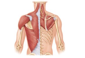
Muscles Crossing the Shoulder Joint
Major Movers of the Arm
The shoulder joint is highly flexible but unstable. The three largest movers are deltoid, pectoralis major, and latissimus dorsi. 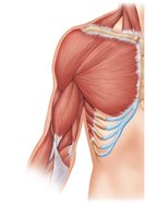
Muscles Crossing the Elbow Joint
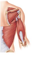
Forearm Extensors and Flexors
Posterior muscles: Extensors (triceps brachii, anconeus).
Anterior muscles: Flexors (biceps brachii, brachialis, brachioradialis).
Muscles of the Forearm
Movements of the Wrist, Hand, and Fingers
Tendons are anchored by flexor and extensor retinacula.
Most forearm muscles originate on the distal humerus.
Movements include flexion, extension, abduction, and adduction.
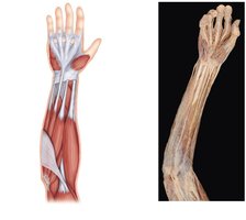
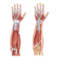
Anterior Flexor Compartment
Includes pronator teres, flexor carpi radialis, palmaris longus, flexor carpi ulnaris, flexor digitorum superficialis.
Innervated by median and ulnar nerves.
Deep Anterior Muscles
Includes flexor pollicis longus, flexor digitorum profundus, pronator quadratus.
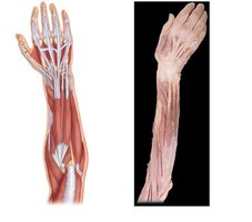
Posterior Extensor Compartment
Includes brachioradialis, extensor carpi radialis longus/brevis, extensor digitorum, extensor carpi ulnaris.
Innervated by the radial nerve.
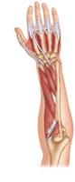
Deep Posterior Muscles
Includes supinator, abductor pollicis longus, extensor pollicis brevis/longus, extensor indicis.
Intrinsic Muscles of the Hand
Fine Movement of the Fingers
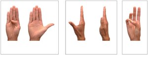
All intrinsic hand muscles are located in the palm and control precise movements such as adduction, abduction, and opposition.
Thenar muscles: Ball of thumb (abductor pollicis brevis, flexor pollicis brevis, opponens pollicis, adductor pollicis).
Hypothenar muscles: Abductor digiti minimi, flexor digiti minimi, opponens pollicis.
Midpalmar muscles: Lumbricals, palmar interossei, dorsal interossei.
Muscles Crossing the Hip and Knee Joints
Thigh and Leg Movements
Anterior muscles: Flex thigh, extend leg at knee.
Posterior muscles: Extend thigh, flex leg.
Adductor muscles: Medial aspect, adduct thigh.
Anterior and Medial Muscles of the Thigh
Origin on pelvis or vertebral column: iliacus, psoas major, sartorius.
Medial compartment: adductor longus, adductor brevis, adductor magnus, pectineus, gracilis.
Quadriceps Femoris
Four heads: rectus femoris, vastus lateralis, vastus medialis, vastus intermedius.
Common insertion at quadriceps tendon; powerful knee extensors.
Posterior Gluteal Muscles and Lateral Rotators
Origin on pelvis or sacrum: gluteus maximus, gluteus medius, gluteus minimus.
Lateral rotators: piriformis, obturator externus/internus, superior/inferior gemellus, quadratus femoris.
Hamstrings
Biceps femoris, semitendinosus, semimembranosus.
Muscles of the Leg
Compartmental Organization
The fascia lata surrounds leg muscles, divides them into three compartments, and aids venous return.
Tendons are held in place by extensor, fibular, and flexor retinacula.
Muscle movement occurs at ankle and intertarsal joints.
Anterior Compartment
Tibialis anterior, extensor digitorum longus, fibularis tertius, extensor hallucis longus.
Lateral Compartment
Fibularis longus, fibularis brevis.
Posterior Compartment
Superficial: triceps surae (gastrocnemius, soleus, plantaris).
Deep: popliteus, flexor digitorum longus, flexor hallucis longus, tibialis posterior.
Intrinsic Muscles of the Foot
Toe Movement and Foot Support
These muscles help flex, extend, abduct, and adduct the toes, and support the arches of the foot.
Single muscle on dorsal aspect: extensor digitorum brevis.
Many muscles on plantar aspect, organized in four layers.
First Layer
Flexor digitorum brevis, abductor hallucis, abductor digiti minimi.
Second Layer
Flexor accessorius, lumbricals.
Third Layer
Flexor hallucis brevis, adductor hallucis, flexor digiti minimi brevis.
Fourth Layer
Plantar and dorsal interossei.
Surface Anatomy
Palpation and Living Anatomy
Palpation is the process of feeling internal structures through the skin, which provides important information about arterial pulses, skeleton, muscles, blood vessels, and the sounds of the heart and lungs. It is also used to determine sites for injections. Additional info: This study guide covers the structure, function, and organization of muscles in the human body, including their development, naming conventions, and compartmentalization. It is suitable for exam preparation in Anatomy & Physiology courses.
