 뒤로
Muscles of the Hip, Thigh, Leg, and Foot: Structure, Function, and Innervation
스터디 가이드 - 스마트 노트
 자료에 맞춘 맞춤형 노트, 핵심 정의, 예시, 맥락을 확장해 제공합니다.
Muscles Crossing the Hip and Knee Joints
Overview of Muscle Groups
Muscles crossing the hip and knee joints are organized into anterior, medial, and posterior groups. Each group is responsible for specific movements of the thigh and leg, and all are enclosed by the fascia lata, the deep fascia of the thigh.
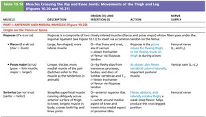
Anterior muscles: Flex the femur at the hip and extend the leg at the knee (foreswing of walking).
Posterior muscles: Extend the thigh and flex the leg (backswing of walking).
Medial (adductor) muscles: Adduct the thigh and have no effect on the leg.
Movements at the Hip Joint
Muscles anchored to the pelvic girdle move the thigh at the hip joint, producing flexion, extension, abduction, adduction, circumduction, and rotation.
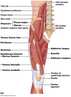
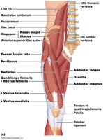
Thigh flexors: Pass in front of the hip joint and include Iliopsoas (iliacus and psoas major), Tensor fasciae latae, Rectus femoris, and are assisted by medial adductors and Sartorius.
Medial (Adductor) Muscles of the Thigh
Adductor Muscles and Their Functions
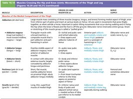
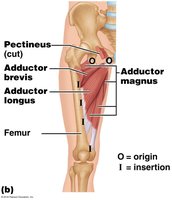
The medial compartment contains adductor muscles that medially rotate the thigh and are used in movements that press the thighs together. These muscles also tilt the pelvis during walking to balance the body’s weight.
Adductor magnus
Adductor longus
Adductor brevis
Pectineus
Gracilis
Anterior Thigh Extensors
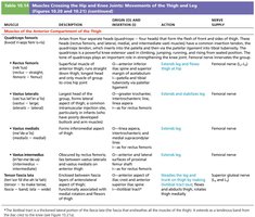
Quadriceps Femoris and Associated Muscles
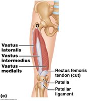
The quadriceps femoris is the prime mover of leg extension and consists of four heads: Rectus femoris, Vastus lateralis, Vastus medialis, and Vastus intermedius. All insert into the quadriceps tendon, which attaches to the patella and then via the patellar ligament to the tibial tuberosity.
Tensor fascia lata: Steadies the leg, flexes, and abducts the thigh.
Quadriceps femoris: Powerful knee extensor used for climbing, running, and jumping.
Posterior Muscles of the Hip and Thigh
Gluteal Muscles and Lateral Rotators
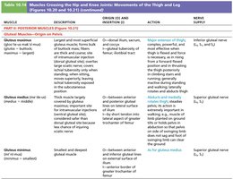
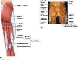
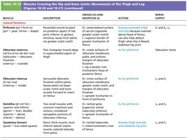
The posterior group includes muscles for thigh extension, abduction, and lateral rotation.
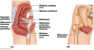
Gluteus maximus: Major thigh extensor and lateral rotator.
Gluteus medius: Abducts and medially rotates thigh.
Gluteus minimus: Medially rotates thigh.
Piriformis, Obturator externus/internus, Gemellus: Lateral rotators of the thigh.
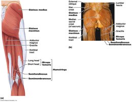
Hamstrings: Muscles of the Posterior Thigh
Structure and Function
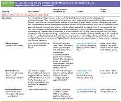
The hamstrings are fleshy muscles of the posterior thigh that cross both the hip and knee joints. They are prime movers of thigh extension and knee flexion and are innervated by the sciatic nerve.
Biceps femoris
Semitendinosus
Semimembranosus
Muscles of the Leg
Compartments and Movements
The deep fascia of the leg is continuous with the fascia lata and segregates the leg into anterior, lateral, and posterior compartments. These compartments contain muscles responsible for movements at the ankle (dorsiflexion and plantar flexion), intertarsal joints (inversion and eversion), and toes (flexion and extension).
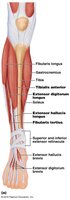
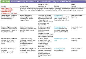
Anterior Compartment Muscles
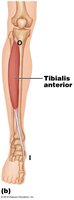
Muscles in the anterior compartment are primary toe extensors and ankle dorsiflexors.
Tibialis anterior
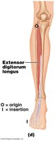
Extensor digitorum longus
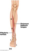
Fibularis tertius
Extensor hallucis longus
Lateral Compartment Muscles
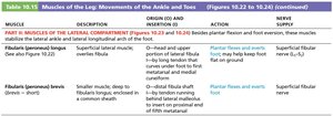
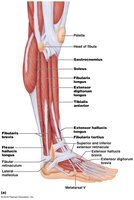
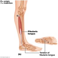
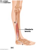
Muscles in the lateral compartment produce plantar flexion and eversion of the foot and stabilize the lateral ankle and arch.
Fibularis longus
Fibularis brevis
Posterior Compartment Muscles
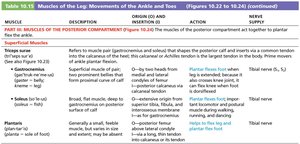
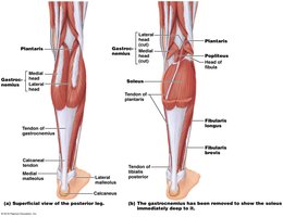
Muscles in the posterior compartment act to plantar flex the ankle and are innervated by the tibial nerve.
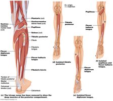
Superficial muscles: Gastrocnemius, Soleus (together called Triceps surae), Plantaris
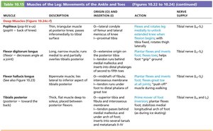
Deep muscles: Popliteus, Flexor digitorum longus, Flexor hallucis longus, Tibialis posterior
Intrinsic Muscles of the Foot
Structure and Function
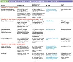
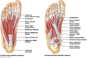
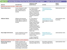
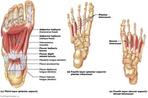
Intrinsic muscles of the foot flex, extend, abduct, and adduct the toes, and support the arches of the foot. The plantar aspect contains four layers of muscles, each contributing to toe movement and arch support.
Dorsal muscle: Extensor digitorum brevis
Plantar muscles: Organized into four layers (superficial to deep)
Summary Tables: Actions of Muscles Acting on the Thigh, Leg, and Foot
Summary of Muscle Actions
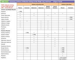
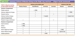
The following tables summarize the actions of muscles acting on the hip, knee, ankle, and toes, including their primary movements and innervation.
Muscle | Hip Flexion | Hip Extension | Abduction | Adduction | Medial Rotation | Lateral Rotation | Knee Flexion | Knee Extension |
|---|---|---|---|---|---|---|---|---|
Iliopsoas | x (PM) | |||||||
Quadriceps femoris | x (PM) | |||||||
Adductor magnus | x (PM) | x | x | |||||
Gluteus maximus | x (PM) | x (PM) | ||||||
Hamstrings | x (PM) | x (PM) |
Muscle | Plantar Flexion | Dorsiflexion | Inversion | Eversion | Toe Flexion | Toe Extension |
|---|---|---|---|---|---|---|
Tibialis anterior | x (PM) | x | ||||
Extensor digitorum longus | x | x | x (PM) | |||
Fibularis longus | x | x | ||||
Gastrocnemius | x (PM) | |||||
Flexor hallucis longus | x | x (PM) |
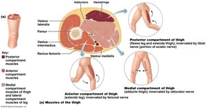
Compartmentalization of Thigh and Leg Muscles
Functional Anatomy
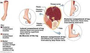
Muscles of the thigh and leg are organized into compartments, each with distinct functions and innervation.
Thigh: Anterior (extends leg), medial (adducts thigh), posterior (flexes leg and extends thigh)
Leg: Anterior (dorsiflexes foot, extends toes), lateral (everts foot), posterior (plantar flexes foot, flexes toes)
Key Terms and Concepts
Prime mover (PM): The muscle primarily responsible for a specific movement.
Fascia lata: Deep fascia enclosing the thigh muscles.
Retinacula: Thickened fascia that holds tendons in place near joints.
Innervation: The nerve supply to a muscle, critical for its function.
Example: Walking
During walking, anterior muscles flex the hip and extend the knee during the foreswing, while posterior muscles extend the hip and flex the knee during the backswing. Medial muscles stabilize the pelvis and adduct the thigh to maintain balance.
Additional info:
The tables and diagrams provide a comprehensive overview of muscle origins, insertions, actions, and innervation, which are essential for understanding musculoskeletal function and clinical assessment in anatomy and physiology.
