 뒤로
Muscles of the Human Body: Structure, Arrangement, and Function
스터디 가이드 - 스마트 노트
 자료에 맞춘 맞춤형 노트, 핵심 정의, 예시, 맥락을 확장해 제공합니다.
Muscle Tissue and Fascicle Arrangement
Patterns of Fascicle Arrangement
The arrangement of muscle fascicles determines the shape and function of skeletal muscles. Fascicles are bundles of muscle fibers, and their organization affects the muscle's range of motion and power.
Circular: Fascicles are arranged in concentric rings, commonly found in sphincter muscles such as the orbicularis oris.
Convergent: Fascicles converge toward a single tendon, as seen in the pectoralis major.
Parallel: Fascicles run parallel to the long axis of the muscle, typical in muscles like the sartorius.
Unipennate: Fascicles insert into only one side of the tendon, as in the extensor digitorum longus.
Bipennate: Fascicles insert into the tendon from both sides, as in the rectus femoris.
Fusiform: Muscle is spindle-shaped with a central belly, as in the biceps brachii.
Multipennate: Fascicles attach to multiple tendons, as in the deltoid.
Example: The arrangement of fascicles in the deltoid muscle allows for a wide range of shoulder movements. 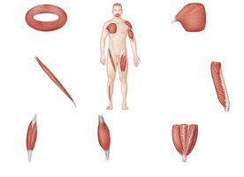
Superficial Muscles of the Body
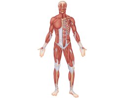
Anterior View
The anterior view of the human body reveals the major superficial muscles responsible for movement and posture. These muscles are grouped by region and function.
Head and Neck: Includes muscles such as the temporalis, masseter, sternocleidomastoid, and platysma.
Thorax: Muscles like the pectoralis major, pectoralis minor, and serratus anterior are involved in movements of the shoulder and rib cage.
Abdomen: The rectus abdominis, external oblique, internal oblique, and transversus abdominis support the trunk and aid in respiration.
Upper Limb: Muscles such as the biceps brachii, triceps brachii, brachialis, and brachioradialis facilitate arm and forearm movements.
Lower Limb: Includes the iliopsoas, rectus femoris, vastus lateralis, vastus medialis, sartorius, adductor longus, gracilis, gastrocnemius, and soleus.
Posterior View
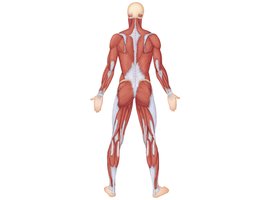
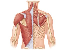
The posterior view highlights muscles involved in extension, rotation, and stabilization of the body.
Neck and Back: Includes the trapezius, latissimus dorsi, rhomboid major, and infraspinatus.
Shoulder and Arm: Muscles such as the deltoid, triceps brachii, and brachialis are visible.
Hip and Thigh: The gluteus maximus, gluteus medius, biceps femoris, adductor magnus, semitendinosus, and semimembranosus are key for locomotion.
Leg: Muscles such as the gastrocnemius, soleus, and fibularis longus are involved in movement of the foot and ankle.
Muscles of the Scalp, Face, and Neck
Facial Muscles
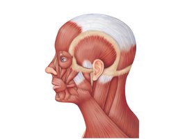
Muscles of the face are responsible for expressions, mastication, and movement of the scalp.
Epicranius: Composed of the frontal and occipital bellies, connected by the galea aponeurotica.
Orbicularis oculi: Closes the eyelids.
Zygomaticus: Raises the corners of the mouth.
Buccinator: Compresses the cheek.
Masseter: Elevates the mandible for chewing.
Platysma: Tenses the skin of the neck.
Neck Muscles
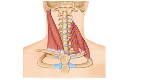
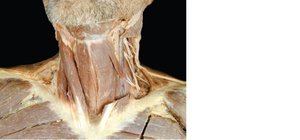
Muscles of the neck facilitate movement of the head and support the cervical spine.
Sternocleidomastoid: Flexes and rotates the head.
Scalene muscles: Assist in flexion and lateral movement of the neck.
Platysma: Superficial muscle that covers the anterior neck.
Muscles of the Abdominal Wall
Structure and Function
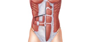
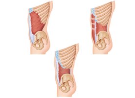
The abdominal wall is composed of several layers of muscles that protect internal organs, support the trunk, and aid in movements such as flexion and rotation.
Rectus abdominis: Runs vertically and is responsible for flexing the vertebral column.
External oblique: Outermost layer, aids in rotation and lateral flexion.
Internal oblique: Lies beneath the external oblique, also assists in rotation and lateral flexion.
Transversus abdominis: Deepest layer, compresses abdominal contents.
Linea alba: Midline connective tissue running vertically.
Inguinal ligament: Formed by the inferior border of the external oblique aponeurosis.
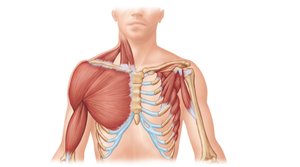
Muscles of the Thorax and Shoulder
Superficial Muscles Acting on the Scapula and Arm
Muscles of the thorax and shoulder are essential for movements of the upper limb and stabilization of the shoulder joint.
Pectoralis major: Adducts and flexes the arm.
Pectoralis minor: Stabilizes the scapula.
Serratus anterior: Protracts the scapula.
Deltoid: Abducts the arm.
Subclavius: Stabilizes the clavicle.
Coracobrachialis: Flexes and adducts the arm.
Muscles Crossing the Shoulder and Elbow Joint
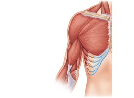
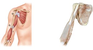
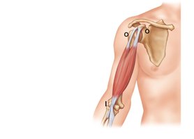
Movements of the Arm and Forearm
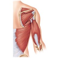
Muscles crossing the shoulder and elbow joints are responsible for flexion, extension, abduction, and rotation of the arm and forearm.
Biceps brachii: Flexes the elbow and supinates the forearm.
Triceps brachii: Extends the elbow.
Brachialis: Flexes the elbow.
Brachioradialis: Flexes the forearm.
Coracobrachialis: Flexes and adducts the arm.
Subscapularis: Medially rotates the arm (part of the rotator cuff).
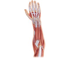
Muscles of the Forearm Acting on the Wrist and Fingers
Anterior Compartment
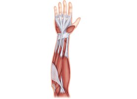
Muscles in the anterior compartment of the forearm are primarily responsible for flexion of the wrist and fingers.
Flexor carpi radialis: Flexes and abducts the wrist.
Palmaris longus: Flexes the wrist.
Flexor carpi ulnaris: Flexes and adducts the wrist.
Flexor digitorum superficialis: Flexes the fingers.
Pronator teres: Pronates the forearm.
Flexor pollicis longus: Flexes the thumb.
Posterior Compartment
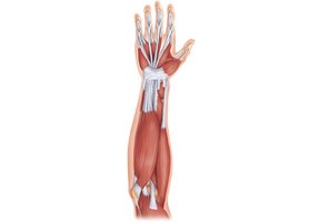
Muscles in the posterior compartment extend the wrist and fingers.
Extensor carpi radialis longus and brevis: Extend and abduct the wrist.
Extensor digitorum: Extends the fingers.
Extensor carpi ulnaris: Extends and adducts the wrist.
Extensor pollicis longus and brevis: Extend the thumb.
Abductor pollicis longus: Abducts the thumb.
Muscles of the Thigh and Leg
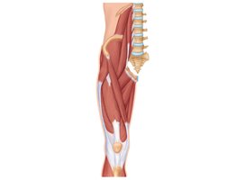
Anterior and Medial Compartment
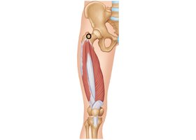
Muscles in the anterior and medial compartments of the thigh promote movements such as extension, flexion, and adduction.
Iliopsoas: Flexes the thigh.
Quadriceps femoris: Extends the knee; includes rectus femoris, vastus lateralis, vastus medialis, and vastus intermedius.
Sartorius: Flexes, abducts, and laterally rotates the thigh.
Adductor longus, magnus, and gracilis: Adduct the thigh.
Posterior Compartment
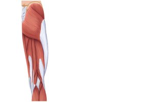
Muscles in the posterior compartment are involved in extension of the hip and flexion of the knee.
Gluteus maximus and medius: Extend and abduct the thigh.
Hamstrings: Include biceps femoris, semitendinosus, and semimembranosus; flex the knee and extend the hip.
Adductor magnus: Adducts the thigh.
Muscles of the Leg and Foot
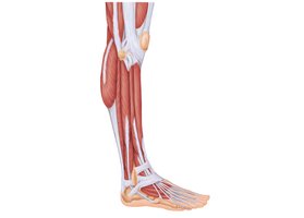
Anterior Compartment
Muscles in the anterior compartment of the leg are responsible for dorsiflexion and extension of the toes.
Tibialis anterior: Dorsiflexes and inverts the foot.
Extensor digitorum longus: Extends the toes.
Extensor hallucis longus: Extends the big toe.
Fibularis longus and tertius: Evert the foot.
Posterior Compartment
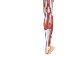
Muscles in the posterior compartment are involved in plantar flexion and flexion of the toes.
Gastrocnemius: Plantar flexes the foot.
Soleus: Plantar flexes the foot.
Flexor digitorum longus: Flexes the toes.
Flexor hallucis longus: Flexes the big toe.
Plantaris: Assists in plantar flexion.
Deep Muscles of the Posterior Compartment
Deep muscles are responsible for fine movements of the toes and stabilization of the foot.
Flexor digitorum longus: Flexes the toes.
Tibialis posterior: Inverts and plantar flexes the foot.
Popliteus: Unlocks the knee joint.
Example: The triceps surae, composed of the gastrocnemius and soleus, is essential for walking and running.
Summary Table: Major Muscle Groups and Their Functions
Region | Major Muscles | Primary Function |
|---|---|---|
Head/Neck | Sternocleidomastoid, Masseter, Platysma | Head movement, mastication, facial expression |
Thorax/Shoulder | Pectoralis major, Deltoid, Serratus anterior | Arm movement, scapular stabilization |
Abdomen | Rectus abdominis, External/Internal oblique | Trunk flexion, rotation, abdominal compression |
Upper Limb | Biceps brachii, Triceps brachii, Brachialis | Flexion/extension of elbow, forearm movement |
Thigh | Quadriceps femoris, Sartorius, Adductors | Knee extension, thigh flexion/adduction |
Leg | Gastrocnemius, Tibialis anterior, Soleus | Plantar/dorsiflexion, toe movement |
Additional info: The notes above expand on the original content by providing definitions, examples, and a summary table for academic completeness.
