 뒤로
Muscles of the Thorax, Abdominal Wall, and Shoulder: Structure and Function Chapter 10 part 2
스터디 가이드 - 스마트 노트
 자료에 맞춘 맞춤형 노트, 핵심 정의, 예시, 맥락을 확장해 제공합니다.
Muscles of Respiration
Deep Muscles of the Thorax: Breathing
The deep muscles of the thorax are essential for the process of breathing, which consists of two phases: inspiration (inhaling) and expiration (exhaling). These muscles alter the size of the thoracic cavity, enabling air movement into and out of the lungs.
Inspiratory Muscles: The diaphragm and external intercostals contract to enlarge the rib cage, increasing thoracic volume and drawing air into the lungs.
Expiratory Muscles: Expiration is primarily passive, resulting from relaxation of inspiratory muscles. However, contraction of the internal intercostals and abdominal muscles can actively decrease thoracic volume, forcing air out.
Diaphragm: This dome-shaped muscle separates the thoracic and abdominal cavities and is the most important muscle for inspiration.
Intercostal Muscles: Located between the ribs, these muscles assist in elevating or depressing the rib cage during breathing.
Example: During vigorous exercise, the internal intercostals and abdominal muscles contract forcefully to enhance expiration.
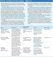
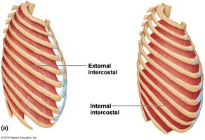
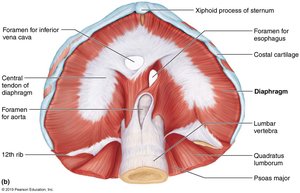
Table: Deep Muscles of the Thorax
Muscle | Description | Origin & Insertion | Action | Nerve Supply |
|---|---|---|---|---|
External intercostals | 11 pairs between ribs; fibers run obliquely downward and forward | Origin: inferior border of rib above; Insertion: superior border of rib below | Pull ribs toward one another to elevate rib cage; aids in inspiration | Intercostal nerves |
Internal intercostals | 11 pairs between ribs; fibers run deep and at right angles to external intercostals | Origin: superior border of rib below; Insertion: inferior border of rib above | Draw ribs together and depress rib cage; aids in forced expiration | Intercostal nerves |
Diaphragm | Broad muscle; forms floor of thoracic cavity | Origin: inferior surface of rib cage, sternum, lumbar vertebrae; Insertion: central tendon | Prime mover of inspiration; flattens on contraction | Phrenic nerves |
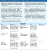
Muscles of the Abdominal Wall
Trunk Movements and Compression of Abdominal Viscera
The abdominal wall is formed by four paired muscles and their connective tissue structures. These muscles are crucial for trunk movement, stabilization, and compression of abdominal contents.
Rectus abdominis: Runs vertically along the midline; responsible for flexing the lumbar region and stabilizing the pelvis.
External obliques: Located on the lateral sides; fibers run diagonally downward.
Internal obliques: Located beneath the external obliques; fibers run diagonally upward.
Transversus abdominis: Deepest muscle; fibers run horizontally.
Fascicle Arrangement: The muscles' fascicles run at different angles, providing strength and flexibility.
Innervation: All are innervated by intercostal nerves.
Actions: Lateral flexion, rotation of the trunk, and compression of abdominal viscera. These actions assist in urination, defecation, childbirth, vomiting, coughing, and screaming.
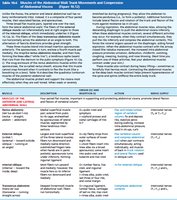
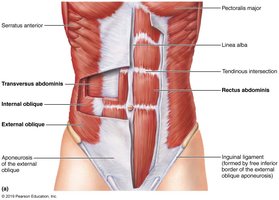
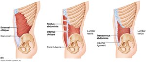

Table: Muscles of the Abdominal Wall
Muscle | Description | Origin & Insertion | Action | Nerve Supply |
|---|---|---|---|---|
Rectus abdominis | Medial muscle; segmented by tendinous intersections | Origin: pubic crest and symphysis; Insertion: xiphoid process and costal cartilages | Flexes and rotates lumbar region; stabilizes pelvis | Intercostal nerves |
External oblique | Largest and most superficial; fibers run downward and medially | Origin: lower eight ribs; Insertion: linea alba, pubic crest, iliac crest | Flexes and rotates vertebral column; compresses abdominal wall | Intercostal nerves |
Internal oblique | Fibers run upward and medially | Origin: lumbar fascia, iliac crest; Insertion: linea alba, pubic crest, last three ribs | Same as external oblique | Intercostal nerves |
Transversus abdominis | Deepest muscle; fibers run horizontally | Origin: inguinal ligament, lumbar fascia, cartilages of last six ribs; Insertion: linea alba, pubic crest | Compresses abdominal contents | Intercostal nerves |
Superficial Muscles of the Anterior and Posterior Thorax
Movements of the Scapula and Arm
These muscles are primarily extrinsic shoulder muscles that act in combination to fix the shoulder girdle and move the scapula, increasing the range of arm movements. They are divided into anterior and posterior groups, each with specific actions.
Actions: Elevation, depression, rotation, lateral and medial movements, protraction, and retraction of the scapula.
Anterior Thorax Muscles: Include pectoralis major, pectoralis minor, serratus anterior, and subclavius.
Posterior Thorax Muscles: Include trapezius, levator scapulae, rhomboids, and latissimus dorsi.
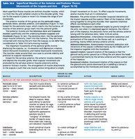
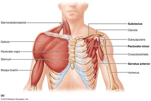
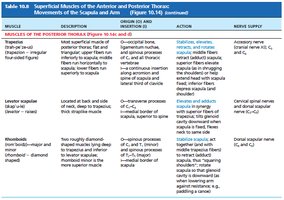
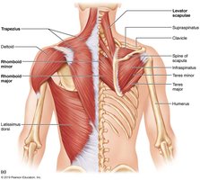
Table: Superficial Muscles of the Anterior and Posterior Thorax
Muscle | Description | Origin & Insertion | Action | Nerve Supply |
|---|---|---|---|---|
Pectoralis major | Large, fan-shaped; covers upper chest | Origin: clavicle, sternum, ribs; Insertion: humerus | Flexes, adducts, and medially rotates arm | Lateral and medial pectoral nerves |
Serratus anterior | Fan-shaped; lies deep to scapula | Origin: ribs; Insertion: scapula | Protracts and holds scapula against thoracic wall | Long thoracic nerve |
Trapezius | Large, triangular; covers back and neck | Origin: occipital bone, vertebrae; Insertion: scapula, clavicle | Elevates, retracts, and rotates scapula | Accessory nerve (CN XI) |
Rhomboids | Rectangular; deep to trapezius | Origin: vertebrae; Insertion: scapula | Stabilizes scapula | Dorsal scapular nerve |
Summary Table: Muscle Actions and Functions
Muscle Group | Main Actions | Key Functions |
|---|---|---|
Thorax (Respiration) | Inspiration, expiration | Ventilation, gas exchange |
Abdominal Wall | Flexion, rotation, compression | Trunk movement, protection, visceral compression |
Shoulder/Thorax | Scapular movement | Arm mobility, shoulder stabilization |
Additional info: The tables and diagrams provided reinforce the anatomical structure, origin, insertion, action, and innervation of each muscle group, which are essential for understanding their physiological roles in movement and stability.
