 뒤로
뒤로Muscular System: Axial and Appendicular Musculature Study Guide
스터디 가이드 - 스마트 노트
 자료에 맞춘 맞춤형 노트, 핵심 정의, 예시, 맥락을 확장해 제공합니다.
The Muscular System: Axial and Appendicular Musculature
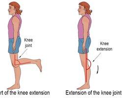
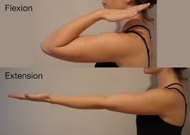
Flexion and Extension
Flexion and extension are fundamental movements at synovial joints, crucial for understanding muscle actions. Flexion decreases the angle between two body parts, while extension increases it. These movements are essential for locomotion and manipulation of objects.
Flexion: Movement that decreases the angle at a joint (e.g., bending the elbow or knee).
Extension: Movement that increases the angle at a joint (e.g., straightening the elbow or knee).
Example: Flexion at the elbow brings the forearm toward the upper arm; extension straightens the arm.
Abduction and Adduction
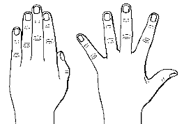
Abduction and adduction describe movements relative to the midline of the body. These terms are commonly used for limbs and fingers.
Abduction: Movement away from the midline (e.g., raising arms or spreading fingers).
Adduction: Movement toward the midline (e.g., lowering arms or bringing fingers together).
Example: Abducting the fingers spreads them apart; adducting brings them together.
Muscles of the Head and Neck
Muscles of Facial Expression and Mastication
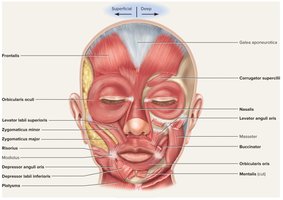
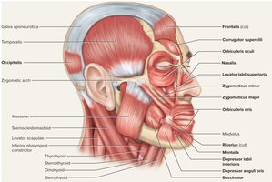
The head and neck contain numerous muscles responsible for facial expression, mastication, and movement of the scalp. The galea aponeurotica (epicranial aponeurosis) is a tough connective tissue layer covering the scalp.
Frontalis: Elevates eyebrows, wrinkles forehead.
Orbicularis oculi: Closes eyelids (winking).
Orbicularis oris: Closes mouth, puckers lips.
Zygomaticus major/minor: Draws angle of lip upward (smiling).
Buccinator: Compresses cheek against teeth.
Masseter: Elevates mandible (chewing).
Galea aponeurotica: Provides attachment for scalp muscles.
Muscles of Head and Neck Region (Lateral View)
Muscles in the lateral view of the head and neck are involved in mastication, facial expression, and movement of the scalp.
Temporalis: Elevates mandible.
Occipitalis: Draws scalp posteriorly.
Sternocleidomastoid: Rotates and flexes the head and neck.
Sternocleidomastoid Muscle
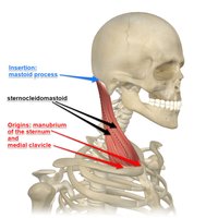
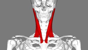
The sternocleidomastoid muscle has two heads (sternal and clavicular) and inserts onto the mastoid process. It is essential for head rotation and flexion.
Origin: Manubrium of sternum and medial clavicle.
Insertion: Mastoid process of temporal bone and superior nuchal line of occipital bone.
Action: Rotates and flexes the head and neck.
Muscles of the Trunk
Intercostal Muscles
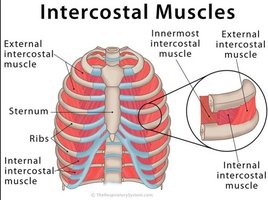
The intercostal muscles are located between the ribs and play a vital role in respiration.
External intercostals: Elevate ribs, expand thoracic cavity, aid in inspiration.
Internal intercostals: Depress and retract ribs, compress thoracic cavity, aid in expiration.
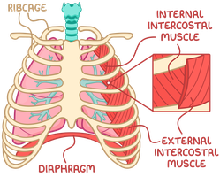
Diaphragm
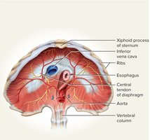
The diaphragm is a muscular dome separating the thoracic and abdominal cavities. It is the primary muscle of respiration.
Contraction: Flattens diaphragm, enlarges thoracic cavity (inspiration).
Relaxation: Raises diaphragm, shrinks thoracic cavity (expiration).
Central tendon: Fibrous tissue where muscle fascicles converge.
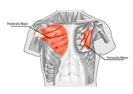
Pectoralis Major and Minor
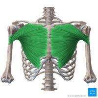
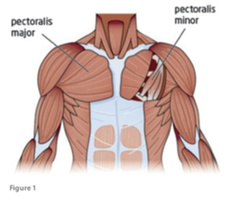
The pectoralis major and pectoralis minor are muscles of the anterior thoracic wall, important for movements of the shoulder and arm.
Pectoralis major: Flexes, adducts, and medially rotates humerus.
Pectoralis minor: Deep to pectoralis major; stabilizes scapula.
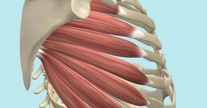
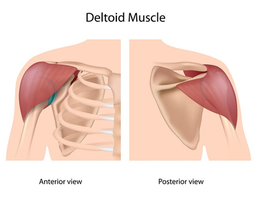
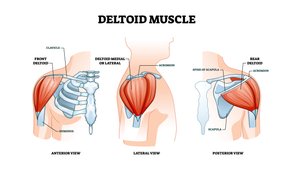
Deltoid and Serratus Anterior
The deltoid muscle is the primary abductor of the arm, while the serratus anterior stabilizes and moves the scapula.
Deltoid: Raises arm laterally.
Serratus anterior: Attaches ribs to medial border of scapula; draws scapula laterally and forward.
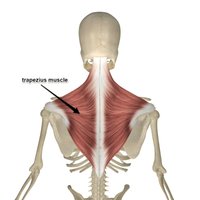
Trapezius
The trapezius muscle stabilizes the scapula and shoulder, and is involved in elevating and depressing the shoulder apex.
Action: Stabilizes, elevates, and depresses the scapula.
Location: Superficial muscle of the upper back.
Latissimus Dorsi
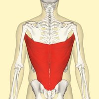
The latissimus dorsi is a large muscle of the back, responsible for adduction and medial rotation of the humerus.
Action: Adducts and medially rotates humerus.
Location: Posterior view, covers lower back.
Rotator Cuff (SITS Muscles)
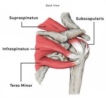
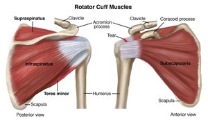
The rotator cuff is a group of four muscles that stabilize the shoulder joint and rotate the arm.
Supraspinatus
Infraspinatus
Teres minor
Subscapularis
Action: Stabilize the shoulder joint and rotate the arm.
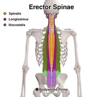
Erector Spinae
The erector spinae is a group of muscles that extend, rotate, and stabilize the spine, maintaining upright posture.
Action: Extension, rotation, stabilization of the spine.
Components: Spinalis, Longissimus, Iliocostalis.
Muscles of the Upper Arm
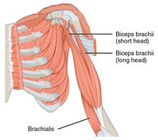
Brachialis and Biceps Brachii
The upper arm contains principal flexors and extensors. The brachialis is the main flexor, while the biceps brachii acts as a synergist.
Brachialis: Principal flexor of the forearm.
Biceps brachii: Synergistic flexor, assists brachialis.
Triceps Brachii
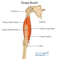
The triceps brachii is the principal extensor of the forearm, with three heads (long, medial, lateral).
Action: Extension of the forearm.
Heads: Long, medial, lateral.
Brachioradialis
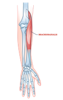
The brachioradialis flexes the forearm at the elbow when the forearm is in a mid-pronated position and assists in forearm rotation.
Action: Flexion of forearm at elbow, assists in pronation and supination.
Muscles of the Forearm
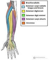
Posterior Forearm Muscles
Muscles in the posterior compartment of the forearm are primarily extensors of the wrist and fingers.
Extensor carpi radialis longus/brevis: Extension and abduction of wrist.
Extensor digitorum: Extension of fingers.
Extensor pollicis: Extension and abduction of thumb.
Extensor carpi ulnaris: Extension and adduction of wrist.
Anterior Forearm Muscles
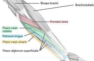
Muscles in the anterior compartment of the forearm are primarily flexors of the wrist and fingers.
Flexor carpi radialis: Flexion and abduction of wrist.
Flexor carpi ulnaris: Flexion and adduction of wrist.
Flexor digitorum superficialis: Flexion of fingers.
Muscles of the Femoral Region
Sartorius
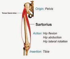
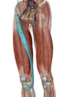
The sartorius is the longest muscle in the body, involved in flexion, abduction, and lateral rotation of the thigh at the hip, and flexion and medial rotation of the leg at the knee.
Origin: Pelvis.
Insertion: Tibia.
Action: Hip flexion, abduction, lateral rotation; knee flexion, medial rotation.
Stabilizes: Pelvis, especially in females.
Medial (Adductor) Compartment of Thigh
The medial compartment contains five primary adductors of the thigh.
Adductor brevis
Adductor longus
Adductor magnus
Gracilis
Pectineus
Mnemonic: 3 Ducks Pecking Grass (Adductor brevis, Adductor longus, Adductor magnus, Gracilis, Pectineus)
Quadriceps Femoris
The quadriceps femoris is the prime mover of knee extension and the most powerful muscle group in the body.
Heads: Rectus femoris, vastus lateralis, vastus medialis, vastus intermedius.
Insertion: Quadriceps (patellar) tendon, patella, patellar ligament, tibial tuberosity.
Hamstring Muscles
The posterior compartment of the thigh contains the hamstring muscles, which are responsible for knee flexion.
Biceps femoris
Semitendinosus
Semimembranosus
Gluteal Muscles
The gluteal muscles are involved in hip extension, abduction, and medial rotation.
Gluteus maximus: Prime hip extensor, forms mass of buttock.
Gluteus medius and minimus: Abduct and medially rotate thigh.
Muscles of the Lower Leg
Posterior Flexor Compartment
The posterior compartment contains muscles responsible for plantar flexion and flexion of the toes.
Gastrocnemius: Plantar flexes foot, flexes knee.
Soleus: Plantar flexes foot.
Triceps surae: Collective name for gastrocnemius and soleus; attaches to calcaneus via the calcaneal (Achilles) tendon.
Flexor digitorum longus: Flexes phalanges.
Flexor hallucis longus: Flexes great toe.
Tibialis posterior: Inverts foot.
Lateral (Fibular) Compartment
The lateral compartment contains muscles that evert the foot.
Fibularis longus
Fibularis brevis
Additional info:
Muscle actions are often paired (agonist/antagonist) to allow controlled movement.
Muscle names often reflect their location, action, or shape (e.g., flexor, extensor, longus, brevis).
Understanding muscle attachments (origin and insertion) is key to predicting their actions.
