 뒤로
뒤로Nervous System Laboratory Study Guide
스터디 가이드 - 스마트 노트
 자료에 맞춘 맞춤형 노트, 핵심 정의, 예시, 맥락을 확장해 제공합니다.
Nervous System Overview
Introduction to the Nervous System
The nervous system is a complex network responsible for coordinating the body's activities by transmitting signals between different parts. It consists of the central nervous system (CNS) and the peripheral nervous system (PNS). - Central Nervous System (CNS): Includes the brain and spinal cord. - Peripheral Nervous System (PNS): Comprises nerves and ganglia outside the CNS.
Neurons
Structure and Function of Neurons
Neurons are the fundamental units of the nervous system, specialized for transmitting electrical and chemical signals. - Cell Body (Soma): Contains the nucleus and organelles; responsible for metabolic activities. - Dendrites: Receive incoming signals from other neurons. - Axon: Conducts electrical impulses away from the cell body. - Axon Hillock: Region where action potentials are initiated. - Axon Terminals: Release neurotransmitters to communicate with other cells. - Synapse: Junction between two neurons or a neuron and another cell. - Presynaptic Cell: Sends the signal. - Postsynaptic Cell: Receives the signal.
Types of Neurons
Neurons are classified based on their structure: - Multipolar Neuron: Multiple dendrites, one axon; most common in CNS. - Bipolar Neuron: One dendrite, one axon; found in sensory organs. - Unipolar Neuron: Single process that splits into two branches; common in PNS.
Myelination and Schwann Cells
Myelination increases the speed of nerve impulse conduction. Schwann cells in the PNS and oligodendrocytes in the CNS produce myelin. - Node of Ranvier: Gaps in myelin sheath where action potentials are regenerated. - Internode: Myelinated segment between nodes. - Neurilemma: Outer layer of Schwann cell membrane. 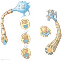
Brain Anatomy
Major Regions of the Brain
The brain is divided into several regions, each with distinct functions. - Cerebrum: Responsible for higher cognitive functions, sensory perception, and voluntary motor actions. - Diencephalon: Includes the thalamus and hypothalamus; relays sensory information and regulates homeostasis. - Brain Stem: Composed of midbrain, pons, and medulla oblongata; controls vital functions. - Cerebellum: Coordinates movement and balance. - Gyri and Sulci: Gyri are ridges, sulci are grooves; increase surface area. 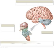
Ventricular System
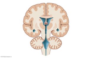
The ventricular system consists of interconnected cavities filled with cerebrospinal fluid (CSF). - Lateral Ventricles: Located in each cerebral hemisphere. - Third Ventricle: Located in the diencephalon. - Fourth Ventricle: Located between the brainstem and cerebellum. - Cerebral Aqueduct: Connects third and fourth ventricles. - Central Canal: Runs through the spinal cord. 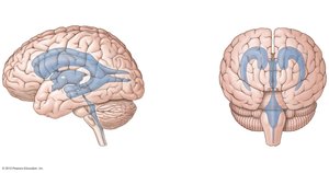
Brain Protection: Meninges and Spaces
Meninges
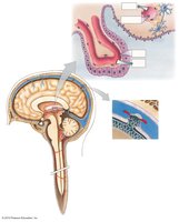
The brain and spinal cord are protected by three layers of connective tissue called meninges. - Dura Mater: Tough, outermost layer. - Arachnoid Mater: Middle, web-like layer. - Pia Mater: Delicate, innermost layer adhering to the brain surface. - Subdural Space: Space between dura mater and arachnoid mater. - Subarachnoid Space: Space between arachnoid mater and pia mater, filled with CSF. 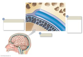
Specialized Structures
- Arachnoid Granulations: Structures that allow CSF to drain into the venous system. - Dural Sinuses: Venous channels located between layers of dura mater. 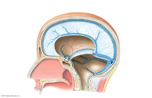
Cerebral Cortex and Functional Areas
Lobes of the Brain
The cerebrum is divided into lobes, each associated with specific functions. - Frontal Lobe: Motor control, problem-solving, speech production. - Parietal Lobe: Sensory perception, spatial reasoning. - Temporal Lobe: Auditory processing, memory. - Occipital Lobe: Visual processing. - Insula: Deep within lateral sulcus; involved in taste and visceral sensation. 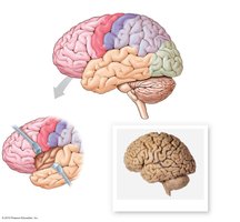
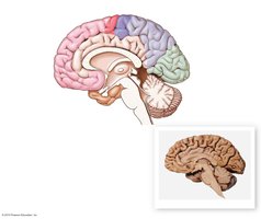
Functional Areas of the Cortex
The cortex contains specialized regions for sensory and motor functions. - Primary Motor Cortex: Controls voluntary movements. - Somatic Motor Association Area: Coordinates learned movements. - Primary Sensory Cortex: Receives somatic sensory information. - Somatic Sensory Association Area: Interprets sensory input. - Visual Cortex: Processes visual information. - Auditory Cortex: Processes auditory information. - Gustatory Cortex: Processes taste. - Olfactory Cortex: Processes smell. 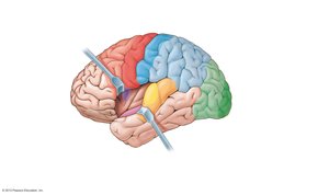
Hemispheric Specialization
The left and right hemispheres of the brain have specialized functions. - Left Hemisphere: Language, mathematical calculation, logic. - Right Hemisphere: Spatial visualization, analysis by touch, creativity. - Corpus Callosum: Connects the two hemispheres, allowing communication. 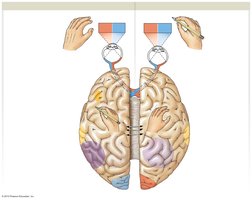
Language Areas
- Broca's Area: Speech production. - Wernicke's Area: Language comprehension. 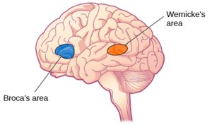
Diencephalon and Brain Stem
Diencephalon
The diencephalon is a central region of the brain containing: - Thalamus: Relay station for sensory information. - Hypothalamus: Regulates homeostasis, endocrine functions, and autonomic control. - Pineal Gland: Produces melatonin, regulates circadian rhythms. 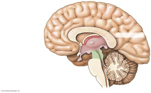
Brain Stem
The brain stem is essential for basic life functions. - Midbrain: Visual and auditory reflexes. - Pons: Relays information between cerebrum and cerebellum; regulates breathing. - Medulla Oblongata: Controls heart rate, blood pressure, and respiration. 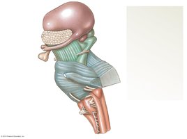
Spinal Cord and Peripheral Nervous System
Spinal Cord Anatomy
The spinal cord is a major pathway for information traveling between the brain and body. - Cervical and Lumbar Enlargements: Regions with increased neural input/output for limbs. - Conus Medullaris: Inferior tip of the spinal cord. - Cauda Equina: Bundle of spinal nerves below the conus medullaris. - Spinal Nerves: Emerge from the spinal cord and branch to various body regions. 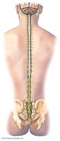
Gray and White Matter
- Gray Matter: Contains neuron cell bodies; organized into horns. - White Matter: Contains myelinated axons; organized into columns. - Dorsal Root: Sensory input to the spinal cord. - Ventral Root: Motor output from the spinal cord.
Dermatomes
Dermatomes are regions of skin innervated by specific spinal nerves. - Clinical Importance: Used to diagnose nerve or spinal cord injuries. 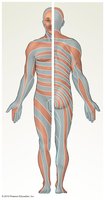
Nerve Plexuses and Peripheral Nerves
Nerve Plexuses
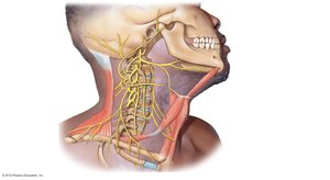
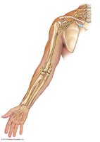
Nerve plexuses are networks of intersecting nerves. - Cervical Plexus: Supplies neck and diaphragm. - Brachial Plexus: Supplies upper limb. - Lumbar Plexus: Supplies anterior thigh. - Sacral Plexus: Supplies posterior thigh, leg, and foot. 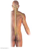
Lumbar and Sacral Plexuses
- Femoral Nerve: Innervates anterior thigh muscles. - Sciatic Nerve: Largest nerve; innervates posterior thigh and leg. 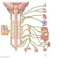
Autonomic Nervous System
Sympathetic Nervous System
The sympathetic division prepares the body for 'fight or flight' responses. - Sympathetic Chain Ganglia: Run alongside the spinal cord. - Preganglionic Neurons: Originate in the spinal cord. - Postganglionic Neurons: Extend to target organs.
Parasympathetic Nervous System
The parasympathetic division promotes 'rest and digest' activities. - Vagus Nerve: Major parasympathetic nerve. - Pelvic Nerves: Innervate lower abdominal organs. - Terminal Ganglia: Located near or within target organs. 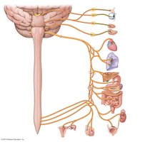
Cranial Nerves
Overview of Cranial Nerves
Cranial nerves emerge directly from the brain and are responsible for sensory and motor functions of the head and neck. - Olfactory (I): Smell - Optic (II): Vision - Oculomotor (III), Trochlear (IV), Abducens (VI): Eye movement - Trigeminal (V): Facial sensation, mastication - Facial (VII): Facial expression, taste - Vestibulocochlear (VIII): Hearing, balance - Glossopharyngeal (IX): Taste, swallowing - Vagus (X): Autonomic control of heart, lungs, digestive tract - Accessory (XI): Neck muscles - Hypoglossal (XII): Tongue movement Additional info: The notes above expand on brief points and organize fragmented content into a comprehensive, structured study guide suitable for college-level Anatomy & Physiology students. All images included are directly relevant to the adjacent content and reinforce key anatomical concepts.
