 뒤로
뒤로Nervous System Microanatomy and Gross Anatomy: Study Notes
스터디 가이드 - 스마트 노트
 자료에 맞춘 맞춤형 노트, 핵심 정의, 예시, 맥락을 확장해 제공합니다.
Nervous System Microanatomy
Structure of a Neuron
The neuron is the fundamental unit of the nervous system, responsible for transmitting electrical and chemical signals. Each neuron consists of several distinct parts:
Cell body (soma): Contains the nucleus and most organelles; site of metabolic activity.
Dendrites: Branch-like extensions that receive signals from other neurons.
Axon: Long, slender projection that transmits electrical impulses away from the cell body.
Axon terminal: End of the axon where neurotransmitters are released.
Axon hillock: Region where the axon originates from the cell body; site of action potential initiation.
Nucleus: Contains genetic material and controls cell function.
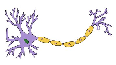
Myelin Sheath and Nodes of Ranvier
The myelin sheath is a fatty layer that insulates axons, increasing the speed of electrical transmission. The Nodes of Ranvier are gaps in the myelin sheath where ion exchange occurs, facilitating rapid signal conduction.
Schwann cells: Produce myelin in the Peripheral Nervous System (PNS).
Oligodendrocytes: Produce myelin in the Central Nervous System (CNS).
Types of Neurons
Neurons are classified based on their structure and function:
Multipolar neurons: Have multiple dendrites and a single axon; most common type in the CNS.
Unipolar neurons: Dendrites and axon are continuous; often found in sensory pathways.
Bipolar neurons: Have one dendrite and one axon; found in special sensory organs (e.g., retina).
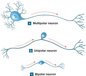
White Matter vs. Gray Matter
White matter consists of myelinated axons, while gray matter contains neuron cell bodies. Their distribution differs between the CNS and PNS:
PNS: Gray matter in ganglia; white matter in nerves.
CNS: Gray matter in cortex and nuclei; white matter in tracts or columns.
Brain Anatomy
Surface Features of the Cerebrum
The cerebrum is characterized by folds and grooves that increase surface area:
Gyrus: Prominent fold or ridge.
Sulcus: Shallow groove.
Fissure: Deep, elongated groove.
Lobes and Functional Areas
The cerebrum is divided into lobes, each with specialized functions:
Frontal lobe: Primary motor cortex, Broca’s area (speech production).
Parietal lobe: Primary sensory cortex.
Occipital lobe: Visual cortex.
Temporal lobe: Auditory cortex, olfactory cortex, Wernicke’s area (language comprehension).
Gustatory cortex: Taste perception.
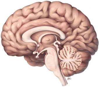
Major Brain Structures
Cerebellum: Coordinates movement and balance.
Medulla oblongata: Controls vital functions (e.g., breathing, heart rate).
Pons: Relays signals between cerebrum and cerebellum.
Midbrain: Processes visual and auditory information.
Corpus callosum: Connects left and right cerebral hemispheres.
Thalamus: Sensory relay station.
Hypothalamus: Regulates homeostasis and endocrine functions.
Pineal gland: Produces melatonin.
Visual Pathways and Associated Structures
The visual system includes the optic nerve, optic chiasm, and optic tract, which transmit visual information from the eyes to the brain.
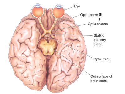
Olfactory Pathways
The olfactory bulb and tract are involved in the sense of smell, transmitting signals from the nasal cavity to the brain.
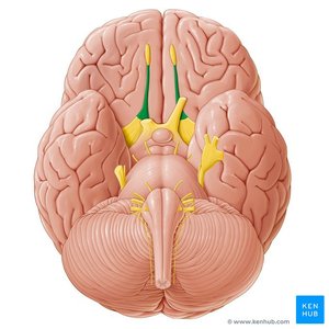
Helpful Mnemonics for Brain Anatomy
Mnemonics can aid in remembering the locations of key brain structures:
Head: thalamus
Cheeks: hypothalamus
Snout: optic chiasm
Ponytail: pineal gland
Neck: midbrain
Belly: pons
Tail: medulla oblongata
Coconut: pituitary gland
Ventricles of the Brain
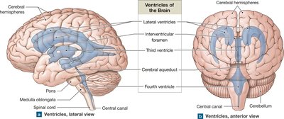
The brain contains interconnected cavities called ventricles, which produce and circulate cerebrospinal fluid (CSF):
Lateral ventricles
Third ventricle
Fourth ventricle
Cerebral aqueduct
Interventricular foramen
Central canal
Choroid plexus: Specialized cells that produce CSF.
Dural Sinuses and Folds
Superior sagittal sinus and inferior sagittal sinus: Venous channels draining blood from the brain.
Falx cerebri: Dural fold separating cerebral hemispheres.
Tentorium cerebelli: Dural fold separating cerebrum from cerebellum.
Spinal Cord Anatomy
Meninges of the Spinal Cord
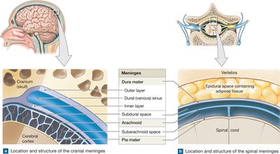
The spinal cord is protected by three layers of connective tissue called meninges:
Dura mater: Tough, outermost layer.
Arachnoid mater: Middle, web-like layer.
Pia mater: Delicate, innermost layer.
Subdural space: Space between dura and arachnoid.
Subarachnoid space: Space between arachnoid and pia, contains CSF.
Spinal Cord Structure
White columns: Contain myelinated axons (posterior, lateral, anterior).
Gray horns: Contain neuron cell bodies (posterior, lateral, anterior).
Gray commissure: Connects left and right sides of gray matter.
Central canal: Contains CSF.
Dorsal root ganglion: Contains sensory neuron cell bodies.
Dorsal (posterior) root: Sensory input.
Ventral (anterior) root: Motor output.
Special Regions and Structures
Conus medullaris: Tapered end of the spinal cord.
Cauda equina: Bundle of nerve roots resembling a horse's tail.
Sympathetic chain ganglia: Chain of nerve bodies running along the spinal cord.
Plexus: Bundle of nerves that innervate specific structures.
Major Nerve Plexuses
Cervical plexus: C1-C4
Brachial plexus: C5-T1
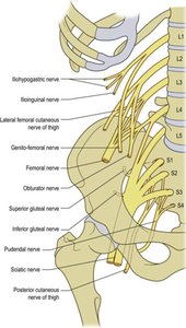
Lumbar plexus: L1-L4
Sacral plexus: L4-S4
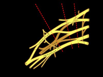
Major Peripheral Nerves
Median nerve
Ulnar nerve
Radial nerve
Musculocutaneous nerve
Axillary nerve
Femoral nerve
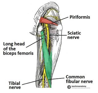
Sciatic nerve
Tibial nerve
Common fibular nerve
Summary Table: Types of Neurons
Type | Structure | Location | Function |
|---|---|---|---|
Multipolar | Multiple dendrites, one axon | CNS | Motor and interneurons |
Unipolar | Dendrites and axon continuous | PNS | Sensory neurons |
Bipolar | One dendrite, one axon | Special senses | Sensory (retina, olfactory) |
Summary Table: Major Nerve Plexuses
Plexus | Spinal Levels | Major Nerves |
|---|---|---|
Cervical | C1-C4 | Phrenic nerve |
Brachial | C5-T1 | Median, ulnar, radial, musculocutaneous, axillary |
Lumbar | L1-L4 | Femoral, obturator |
Sacral | L4-S4 | Sciatic, pudendal |
Additional info: Academic context was added to clarify neuron types, brain regions, and nerve plexuses. Tables were inferred for clarity and completeness.
