 뒤로
Nervous Tissue: Structure, Function, and Electrophysiology
스터디 가이드 - 스마트 노트
 자료에 맞춘 맞춤형 노트, 핵심 정의, 예시, 맥락을 확장해 제공합니다.
Overview of the Nervous System
Functions of the Nervous System
The nervous system is responsible for detecting stimuli, integrating information, and coordinating responses throughout the body. It is divided into two main anatomical divisions: the Central Nervous System (CNS) and the Peripheral Nervous System (PNS).
Sensory Functions: Detection of sensations from inside and outside the body (e.g., pain, temperature, pressure).
Integrative Functions: Analysis and interpretation of sensory input, decision-making, and determination of appropriate responses (primarily in the CNS).
Motor Functions: Stimulation of muscle contraction or gland secretion to produce a response.
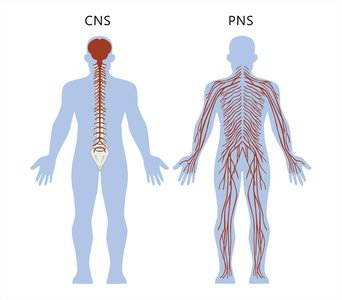
Anatomical Divisions
Central Nervous System (CNS): Consists of the brain and spinal cord. Responsible for processing sensory data, higher brain functions (intelligence, memory, emotion), and transmitting motor commands.
Peripheral Nervous System (PNS): Includes all nervous tissue outside the CNS. Coordinates communication between the CNS and the rest of the body, connecting to muscles, glands, and sensory receptors.
Subdivisions of the PNS
Sensory (Afferent) Division: Transmits sensory information from receptors to the CNS. Includes somatic receptors (muscles, bones, joints, skin), special senses (vision, taste, smell, hearing, balance), and visceral receptors (organs).
Motor (Efferent) Division: Transmits commands from the CNS to effectors (muscles and glands). Subdivided into:
Somatic Nervous System: Voluntary control of skeletal muscles.
Autonomic Nervous System: Involuntary control of smooth muscle, cardiac muscle, and glands. Includes sympathetic ("fight or flight"), parasympathetic ("rest and digest"), and enteric (GI tract control) divisions.
Histology of Nervous Tissue
Cell Types in Nervous Tissue
Nervous tissue is composed of two main types of cells: neurons and neuroglia (glial cells).
Neurons: Excitable cells that generate and conduct action potentials. They are the functional units of the nervous system.
Neuroglia: Supportive cells that provide structural support, growth factors, and insulation for neurons. They are more numerous than neurons and can divide.
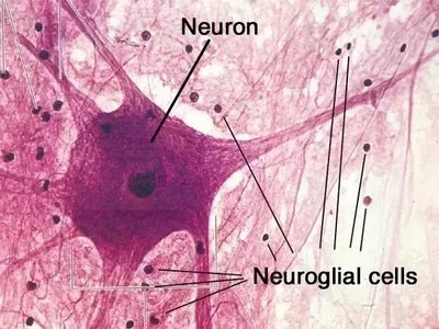
Neurons vs. Nerves
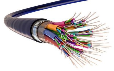
Neuron: A single nerve cell that can be very long. It may be sensory (sending action potentials to the CNS) or motor (carrying action potentials away from the CNS).
Nerve: A bundle of hundreds to thousands of individual neurons, which may contain both sensory and motor fibers.
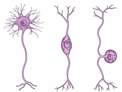
Structural Classification of Neurons
Neurons are classified based on the number and arrangement of their processes:
Multipolar: Several dendrites and one axon; most common type; all motor neurons are multipolar.
Bipolar: One main dendrite and one axon; found in the retina, inner ear, and olfactory epithelium.
Unipolar: Single process with the cell body off to the side; always sensory neurons, often forming ganglia.
Structure of a Typical Neuron
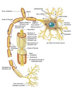
Dendrites: Receive information and create graded potentials. Short, highly branched, and unmyelinated.
Cell Body (Soma): Contains the nucleus and organelles; controls metabolic functions.
Axon: Conducts action potentials away from the cell body; may be myelinated or unmyelinated.
Axon Terminals: End bulbs that connect to muscles, glands, or other neurons; contain neurotransmitters.
Functional Classification of Neurons
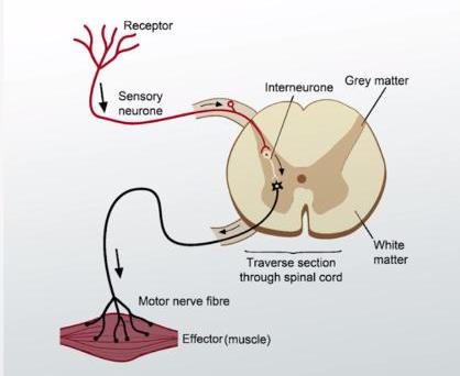
Sensory (Afferent) Neurons: Transmit sensory information to the CNS; generally unipolar or bipolar.
Motor (Efferent) Neurons: Carry instructions from the CNS to effectors; always multipolar.
Interneurons: Located entirely within the CNS; connect sensory and motor neurons; involved in memory, planning, and learning.
Neuroglial (Glial) Cells
Types of Neuroglia
Neuroglia are classified based on their location and function:
Central Nervous System (CNS) | Peripheral Nervous System (PNS) |
|---|---|
Astrocytes | Satellite cells |
Oligodendrocytes | Schwann cells |
Microglia | |
Ependymal cells |
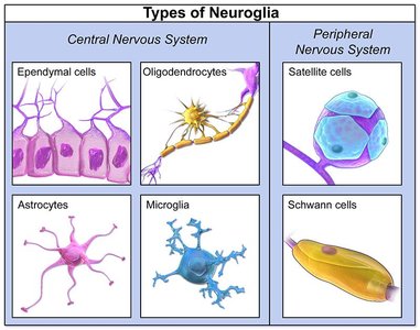
Astrocytes (CNS): Star-shaped, most abundant; anchor neurons and blood vessels, regulate ion concentrations, and help form the blood-brain barrier.
Oligodendrocytes (CNS): Form myelin sheaths around axons in the CNS, increasing conduction speed.
Microglia (CNS): Act as macrophages, providing immune defense and removing debris.
Ependymal Cells (CNS): Line ventricles and central canal; produce and circulate cerebrospinal fluid (CSF).
Schwann Cells (PNS): Form myelin sheaths around axons in the PNS; aid in regeneration of damaged fibers.
Satellite Cells (PNS): Surround neuron cell bodies in ganglia; support and regulate the environment around neurons.
Gray and White Matter
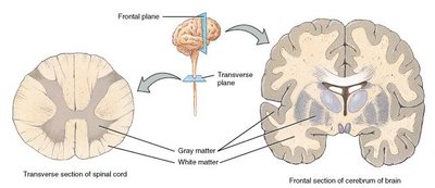
White Matter: Composed of myelinated axons; appears white due to myelin.
Gray Matter: Contains neuron cell bodies, dendrites, unmyelinated axons, and neuroglia; appears gray.
In the spinal cord, gray matter forms an H-shaped core surrounded by white matter; in the brain, gray matter forms a thin outer shell (cortex).
Electrophysiology of Neurons
Ion Channels and Membrane Potential
The movement of ions across the plasma membrane creates electrical gradients, which are the basis for nerve impulses.
Leakage Channels: Randomly open and close, allowing passive diffusion of ions.
Gated Channels: Open in response to specific stimuli:
Voltage-gated: Open in response to changes in membrane potential.
Ligand-gated: Open in response to binding of a chemical messenger (e.g., neurotransmitter).
Mechanically-gated: Open in response to physical deformation of the membrane.
Resting Membrane Potential
The resting membrane potential is the electrical potential difference across the membrane of a neuron at rest, typically around -70 mV (inside negative relative to outside). This is maintained by:
Unequal distribution of ions (Na+, K+, Cl-, proteins).
Leakage channels (more K+ leaks out than Na+ leaks in).
Na+/K+ ATPase pump (actively transports 3 Na+ out and 2 K+ in per ATP hydrolyzed).
Equation for the Nernst potential (for a single ion):
Additional info: R = gas constant, T = temperature, z = charge of ion, F = Faraday's constant.
Graded Potentials vs. Action Potentials
Graded Potentials: Small, localized changes in membrane potential; can be excitatory (EPSP) or inhibitory (IPSP); occur mainly in dendrites and cell body.
Action Potentials: Large, rapid changes in membrane potential that propagate along the axon; all-or-none response; initiated if threshold (-55 mV) is reached at the axon hillock.
Action Potential Phases
Depolarization: Voltage-gated Na+ channels open, Na+ rushes in, membrane potential becomes positive.
Repolarization: Voltage-gated K+ channels open, K+ rushes out, membrane potential returns to negative.
Hyperpolarization: Membrane potential temporarily becomes more negative than resting potential.
Return to Resting Potential: Ion gradients restored by Na+/K+ ATPase.
Propagation of Action Potentials
Continuous Conduction: Occurs in unmyelinated axons; action potential moves along every segment of the membrane; relatively slow.
Saltatory Conduction: Occurs in myelinated axons; action potential "jumps" from node to node (nodes of Ranvier); much faster.
Classification of Axons by Conduction Speed
Type | Diameter | Myelination | Speed | Location |
|---|---|---|---|---|
Type A | 5-20 µm | Always myelinated | ~120 m/s | Sensory/motor to skeletal muscle |
Type B | 2-3 µm | Sometimes myelinated | ~15 m/s | Sensory from organs |
Type C | 0.5-1.5 µm | Unmyelinated | 0.5-2 m/s | Pain, temperature, pressure |
Synapses and Neurotransmitters
Chemical Synapses
Most communication between neurons occurs at chemical synapses, where neurotransmitters are released from the presynaptic neuron and bind to receptors on the postsynaptic neuron.
Neurotransmitters can be excitatory or inhibitory and are quickly removed from the synaptic cleft by enzymes, reuptake, or diffusion.
Neural Circuits
Divergence: One presynaptic neuron synapses with multiple postsynaptic neurons (common in sensory pathways).
Convergence: Multiple presynaptic neurons synapse with a single postsynaptic neuron.
Clinical Correlations
Multiple Sclerosis (MS): Autoimmune disease where myelin sheaths are attacked, disrupting action potentials.
Parkinson's Disease: Neurodegenerative disorder affecting dopamine-producing cells, leading to motor control issues.
Alzheimer's Disease: Progressive loss of neurons in the cerebral cortex, resulting in memory and cognitive decline.
Summary Table: Types of Neuroglia
Type | Location | Function |
|---|---|---|
Astrocytes | CNS | Support, blood-brain barrier, regulate ions |
Oligodendrocytes | CNS | Form myelin sheaths |
Microglia | CNS | Immune defense, phagocytosis |
Ependymal cells | CNS | Produce/circulate CSF |
Schwann cells | PNS | Form myelin sheaths, regeneration |
Satellite cells | PNS | Support neuron cell bodies |
