 뒤로
neuro 8
스터디 가이드 - 스마트 노트
 자료에 맞춘 맞춤형 노트, 핵심 정의, 예시, 맥락을 확장해 제공합니다.
Brain Structure and Function
Diencephalon
The diencephalon is a central region of the brain located between the brain stem and the cerebrum. It plays a crucial role in sensory relay, homeostasis, and endocrine regulation.
Thalamus: Acts as the main sensory relay center, processing and transmitting sensory information to the cerebral cortex.
Hypothalamus: Regulates homeostasis through neural and endocrine outputs, controlling body temperature, hunger, thirst, and circadian rhythms.
Pineal gland: Secretes melatonin, which regulates sleep-wake cycles.
Pituitary gland: Divided into anterior (endocrine tissue) and posterior (neural tissue) sections, it releases hormones that control growth, metabolism, and reproduction.
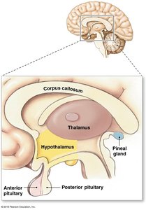
Pineal Gland and Melatonin
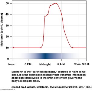
The pineal gland produces melatonin, a hormone that regulates circadian rhythms and sleep. Melatonin secretion increases at night and decreases during the day, acting as a chemical messenger for light-dark cycles.
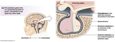
Pituitary Gland Structure and Function
The pituitary gland is connected to the hypothalamus by the infundibulum. The posterior pituitary is an extension of neural tissue, while the anterior pituitary is a true endocrine gland.
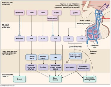
Hypothalamic-Hypophyseal Portal System
The hypothalamus controls the anterior pituitary via the hypophyseal portal system, releasing hormones that regulate growth, metabolism, and reproduction. The anterior pituitary secretes hormones such as prolactin, TSH, ACTH, GH, FSH, and LH, which act on various endocrine and non-endocrine targets.
Cerebrum
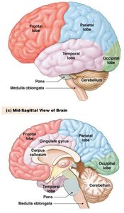
The cerebrum is the largest and most distinctive part of the brain, responsible for higher processing and intelligence. It consists of gray matter (cerebral cortex, basal ganglia, limbic system) and white matter (tracts).
Two hemispheres: Divided into four lobes (frontal, parietal, temporal, occipital), connected by the corpus callosum.
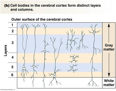
Gray matter: Contains cell bodies and is involved in processing information.
White matter: Composed of myelinated axons that transmit signals between regions.
Basal Ganglia (Basal Nuclei)
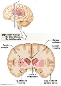
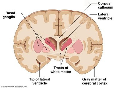
The basal ganglia are three nuclei (globus pallidus, putamen, caudate nucleus) that regulate the initiation and termination of movement. They receive input from the cerebral cortex and provide output to motor areas.
Limbic System
The limbic system, known as the "emotional brain," plays a primary role in emotions, learning, and memory. Major components include the cingulate gyrus, amygdala, and hippocampus.
Cerebral Cortex
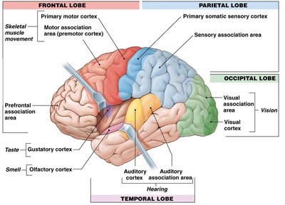
The cerebral cortex is the outermost layer of the cerebrum and serves as the integrating center for the CNS. It is functionally divided into three specializations:
Sensory areas: Translate sensory input into perception.
Motor areas: Direct skeletal muscle movement.
Association areas: Responsible for complex integrative functions such as memory, emotions, reasoning, will, judgment, personality, and intelligence.
Sulci vs Gyri
Sulci are grooves or fissures on the cerebral surface, while gyri are raised ridges. These structures increase the surface area for cortical processing.
Cerebral Lateralization
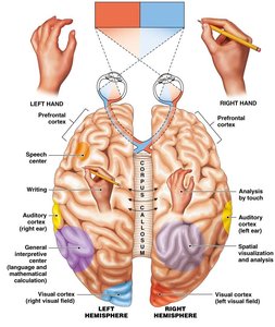
Functional areas are distributed asymmetrically between the two hemispheres, a phenomenon known as cerebral lateralization. For example, language is typically dominant in the left hemisphere, while spatial abilities are more prominent in the right.
Cortical Plasticity
The cortex can undergo plasticity, meaning it can adapt and reorganize in response to experience or injury.
Sensory Physiology
General Properties of Sensory Systems
Sensory systems provide information about the external and internal environment. Sensory neurons possess transducers (receptors) that convert physical stimuli into intracellular signals, typically changes in membrane potential.
Sensory Receptors
Sensory receptors are specialized cells or neurons that detect specific stimuli. Types include:
Chemoreceptors: Detect chemical changes (e.g., blood glucose, pH).
Nociceptors: Detect pain and tissue damage.
Proprioceptors: Sense body position and movement.
Auditory receptors: Hair cells in the ear detect sound.
Balance receptors: Cupula in the vestibular system senses equilibrium.
Vision receptors: Rods and cones in the retina detect light.
Thermal receptors: Detect temperature changes.
Types of Sensory Receptors (Table)
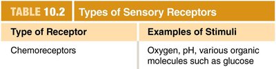
The following table summarizes the main types of sensory receptors and their stimuli:
Type of Receptor | Examples of Stimuli |
|---|---|
Chemoreceptors | Oxygen, pH, various organic molecules such as glucose |
Mechanoreceptors | Pressure (baroreceptors), cell stretch (osmoreceptors), vibration, acceleration, sound |
Thermoreceptors | Varying degrees of heat |


Transduction and Receptor Potentials
Physical stimuli are transduced into receptor potentials (graded potentials). If these reach threshold, they induce action potentials. Mechanically gated channels convert mechanical stimuli into electrical signals.
Somatosensory Neurons and Receptive Fields
Somatosensory neurons bring information to the CNS. Each sensory neuron is activated by stimuli within a specific receptive field, a physical area where the stimulus can be detected.
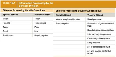
Stimulus Processing (Table)
The following table compares conscious and subconscious stimulus processing:
Stimulus Processing Usually Conscious | Stimulus Processing Usually Subconscious |
|---|---|
Special Senses: Vision, Hearing, Taste, Smell, Equilibrium Somatic Senses: Touch, Temperature, Pain, Itch, Proprioception | Somatic Stimuli: Muscle length and tension, Proprioception Visceral Stimuli: Blood pressure, Distension of gastrointestinal tract, Blood glucose concentration, Internal body temperature, Osmolarity of body fluids, Lung inflation, pH of cerebrospinal fluid, pH and oxygen content of blood |
CNS Integration of Sensory Information
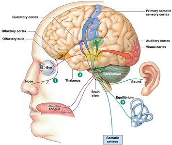
Almost all special and somatic sensory information is routed through the thalamus. Special senses have dedicated cortical regions, while somatic senses are integrated in the primary somatosensory cortex. Visceral sensory information is integrated in the brainstem and spinal cord.
Olfactory Pathways
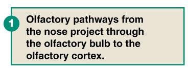
Olfactory pathways from the nose project through the olfactory bulb to the olfactory cortex.
Additional info:
Plasticity of the cortex allows for adaptation and recovery after injury.
Basal ganglia dysfunction is associated with movement disorders such as Parkinson's disease.
Limbic system dysfunction can affect emotional regulation and memory.
