 뒤로
Osseous Tissue and Bone Physiology Study Guide
스터디 가이드 - 스마트 노트
 자료에 맞춘 맞춤형 노트, 핵심 정의, 예시, 맥락을 확장해 제공합니다.
Q1. List and describe the four major functions of bone tissue.
Background
Topic: Bone Physiology
This question tests your understanding of the essential roles that bone tissue plays in the human body, which are foundational to anatomy and physiology.
Key Terms:
Support
Protection
Movement
Mineral Storage
Blood Cell Production
Step-by-Step Guidance
Recall the main physiological functions of bone tissue. Think about how bones contribute to the structure and function of the body.
For each function, write a brief description explaining its importance. For example, consider how bones protect internal organs or serve as sites for blood cell formation.
Try to connect each function to a real-life example or a specific bone in the body.
Try solving on your own before revealing the answer!
Final Answer:
Support: Bones provide a framework for the body, supporting soft tissues and giving shape.
Protection: Bones protect vital organs (e.g., skull protects the brain, ribs protect the heart and lungs).
Movement: Bones act as levers for muscles, enabling movement.
Mineral Storage: Bones store minerals such as calcium and phosphorus, which can be released into the bloodstream as needed.
Blood Cell Production: The bone marrow produces red and white blood cells (hematopoiesis).
Each function is essential for maintaining homeostasis and overall health.
Q2. Complete the table describing the two types of osseous tissue: Compact Bone and Spongy Bone.
Background
Topic: Osseous Tissue Microanatomy
This question tests your knowledge of the structural differences between compact and spongy bone, including their functional units and locations within a long bone.
Key Terms:
Compact Bone (Cortical Bone)
Spongy Bone (Trabecular Bone)
Osteon
Trabeculae
Red Bone Marrow
Step-by-Step Guidance
Identify the functional unit of each bone tissue type (e.g., osteon for compact bone).
Recall where each tissue type is located within a long bone (e.g., diaphysis vs. epiphysis).
Describe what each tissue contains, such as marrow or blood vessels.
Fill in the table with these details, making sure to distinguish between compact and spongy bone.

Try solving on your own before revealing the answer!
Final Answer:
Compact Bone: Functional unit is the osteon; located in the diaphysis (shaft) of long bones; contains blood vessels and nerves.
Spongy Bone: Functional unit is the trabeculae; located in the epiphysis (ends) of long bones; contains red bone marrow.
Red Bone Marrow: Found within the spaces of spongy bone; responsible for hematopoiesis.
This distinction is important for understanding bone strength and function.
Q3. Fill out the table below describing the locations and functions of these tissues: Ligament, Tendon, Articular Cartilage, Periosteum.
Background
Topic: Associated Tissues of Bone Organs
This question tests your ability to identify and describe the connective tissues associated with bones and their roles in the musculoskeletal system.
Key Terms:
Ligament: Connects bone to bone
Tendon: Connects muscle to bone
Articular Cartilage: Covers joint surfaces
Periosteum: Membrane covering bone
Step-by-Step Guidance
Recall the location and function of each tissue type. For example, ligaments are found at joints, tendons at muscle attachments.
Write a brief description for each tissue, focusing on its role in movement or protection.
Think about how these tissues interact with bone to facilitate movement and stability.
Try solving on your own before revealing the answer!
Final Answer:
Ligament: Connects bone to bone; stabilizes joints.
Tendon: Connects muscle to bone; transmits force for movement.
Articular Cartilage: Covers joint surfaces; reduces friction and absorbs shock.
Periosteum: Covers the outer surface of bone; contains nerves and blood vessels, aids in bone growth and repair.
These tissues are essential for joint function and bone health.
Q4. Identify, label, and describe classification of bone (and provide an example for) based on shape, anatomical structure, microscopic anatomy, and location.
Background
Topic: Bone Classification
This question tests your ability to classify bones using different criteria and to provide examples for each classification.
Key Terms:
Shape: Long, short, flat, irregular
Anatomical Structure: Compact vs. spongy
Microscopic Anatomy: Osteons, trabeculae
Location: Axial vs. appendicular skeleton
Step-by-Step Guidance
List the main bone shape categories and give an example for each (e.g., femur for long bone).
Describe how bones are classified by anatomical structure and microscopic anatomy.
Identify the location of each bone type within the skeleton.
Label the indicated parts of a long bone using an image if provided.
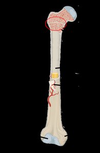
Try solving on your own before revealing the answer!
Final Answer:
Shape: Long (femur), short (carpals), flat (sternum), irregular (vertebrae).
Anatomical Structure: Compact bone forms the outer layer; spongy bone is inside.
Microscopic Anatomy: Osteons in compact bone; trabeculae in spongy bone.
Location: Axial skeleton (skull, vertebrae); appendicular skeleton (limbs).
Classification helps in understanding bone function and structure.
Q5. Define the following bone related anatomical terms and identify their meanings and examples.
Background
Topic: Bone Markings and Terminology
This question tests your knowledge of anatomical terms used to describe bone features and their functional significance.
Key Terms:
Foramen: Hole
Fossa: Depression
Process: Projection
Condyle: Rounded articular surface
Trochanter, Tubercle, Epicondyle, Spine, Facet, Head, Neck, Sinus
Step-by-Step Guidance
List each term and write its meaning (e.g., foramen = hole).
Identify whether the marking is a depression, projection, or hole.
Provide an example of a bone that has each marking.
Try solving on your own before revealing the answer!
Final Answer:
Foramen: Hole (e.g., foramen magnum in the skull)
Fossa: Depression (e.g., olecranon fossa in the humerus)
Process: Projection (e.g., mastoid process in the temporal bone)
Condyle: Rounded articular surface (e.g., femoral condyle)
Other terms: Trochanter (femur), Tubercle (humerus), Epicondyle (humerus), Spine (scapula), Facet (vertebrae), Head (femur), Neck (femur), Sinus (frontal bone)
These markings are important for muscle attachment and joint articulation.
Q6. Identify the functions and locations of the following cells: Osteogenic cell, Osteoblast, Osteocyte, Osteoclast.
Background
Topic: Cellular Components of Bone
This question tests your understanding of the different cell types involved in bone formation, maintenance, and remodeling.
Key Terms:
Osteogenic cell: Stem cell
Osteoblast: Bone-forming cell
Osteocyte: Mature bone cell
Osteoclast: Bone-resorbing cell
Step-by-Step Guidance
Recall the location of each cell type within bone tissue (e.g., osteogenic cells in periosteum).
Describe the function of each cell type (e.g., osteoblasts build bone).
Think about how these cells interact during bone growth and remodeling.
Try solving on your own before revealing the answer!
Final Answer:
Osteogenic cell: Located in periosteum and endosteum; differentiate into osteoblasts.
Osteoblast: Found on bone surfaces; synthesize new bone matrix.
Osteocyte: Embedded in bone matrix; maintain bone tissue.
Osteoclast: Found at sites of bone resorption; break down bone matrix.
These cells are essential for bone health and adaptation.
Q7. Describe the functions of the following hormones as they apply to bone: Calcitonin, Thyroxine, Parathyroid Hormone (PTH), hGH, Cortisol, Testosterone/Estrogen.
Background
Topic: Hormonal Regulation of Bone
This question tests your understanding of how hormones influence bone growth, remodeling, and calcium homeostasis.
Key Terms:
Calcitonin: Lowers blood calcium
Thyroxine: Stimulates bone growth
PTH: Raises blood calcium
hGH: Growth hormone
Cortisol: Stress hormone
Testosterone/Estrogen: Sex hormones
Step-by-Step Guidance
Recall which gland releases each hormone and its effect on bone tissue.
Describe how each hormone influences bone growth or calcium levels.
Think about the physiological consequences of hormone imbalances.
Try solving on your own before revealing the answer!
Final Answer:
Calcitonin: Released by thyroid; decreases blood calcium by inhibiting osteoclasts.
Thyroxine: Released by thyroid; stimulates bone growth.
PTH: Released by parathyroid; increases blood calcium by stimulating osteoclasts.
hGH: Released by pituitary; promotes bone growth.
Cortisol: Released by adrenal; can decrease bone formation.
Testosterone/Estrogen: Released by gonads; promote bone growth and maintenance.
Hormones are key regulators of bone health and development.
Q8. Draw/explain a calcium homeostasis negative feedback loop. Include all the components discussed in class.
Background
Topic: Calcium Homeostasis
This question tests your understanding of how the body maintains stable blood calcium levels through negative feedback mechanisms involving hormones and organs.
Key Terms:
Negative Feedback Loop
Stimulus, Receptor, Control Center, Effector, Response
Calcitonin, PTH
Step-by-Step Guidance
Identify the stimulus (e.g., change in blood calcium levels).
Describe the role of receptors and control centers (e.g., parathyroid gland).
Explain the effector organs and their responses (e.g., bone, kidneys).
Draw or outline the feedback loop, showing hormone release and effects.
Try solving on your own before revealing the answer!
Final Answer:
Stimulus: Low blood calcium triggers parathyroid hormone release.
Receptor: Parathyroid gland senses calcium levels.
Control Center: Parathyroid gland releases PTH.
Effector: Bone (osteoclasts), kidneys, intestines.
Response: Increased blood calcium.
Calcitonin is released when calcium is high, lowering levels.
This loop maintains calcium homeostasis in the body.
Q9. Describe the following and give an example of a bone created by each method: Intramembranous ossification and Endochondral ossification.
Background
Topic: Bone Development
This question tests your understanding of the two main processes by which bones form during development.
Key Terms:
Intramembranous Ossification
Endochondral Ossification
Step-by-Step Guidance
Describe the process of intramembranous ossification and identify a bone formed by this method.
Describe the process of endochondral ossification and identify a bone formed by this method.
Compare the two methods and their significance in skeletal development.
Try solving on your own before revealing the answer!
Final Answer:
Intramembranous Ossification: Bone develops directly from mesenchyme; example: flat bones of the skull.
Endochondral Ossification: Bone develops from cartilage model; example: long bones like the femur.
Both processes are essential for proper skeletal formation.
Q10. Bone Growth Questions: How do bones grow in length? What is appositional bone growth?
Background
Topic: Bone Growth and Development
This question tests your understanding of the mechanisms by which bones increase in size during childhood and adolescence.
Key Terms:
Epiphyseal Plate
Appositional Growth
Step-by-Step Guidance
Describe how bones grow in length at the epiphyseal plate.
Explain the process of appositional growth and how it increases bone diameter.
Think about the cellular activities involved in each type of growth.
Try solving on your own before revealing the answer!
Final Answer:
Length: Bones grow in length by cartilage growth at the epiphyseal plate, followed by ossification.
Appositional Growth: Bones grow in diameter by new bone formation at the periosteum.
These processes are crucial for normal skeletal development.
Q11. Classify the following fracture types based on the images provided.
Background
Topic: Bone Fractures
This question tests your ability to identify and classify different types of bone fractures using visual cues.
Key Terms:
Fracture Types: Transverse, Oblique, Spiral, Comminuted, Greenstick, Impacted
Step-by-Step Guidance
Examine each image and note the direction and pattern of the fracture.
Recall the definitions of common fracture types.
Match each image to the correct fracture classification.
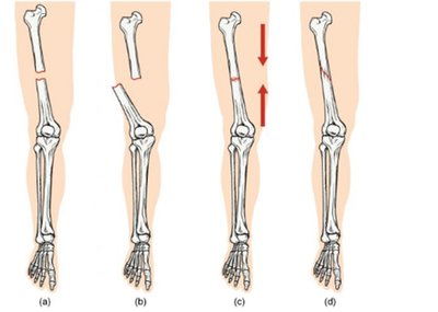
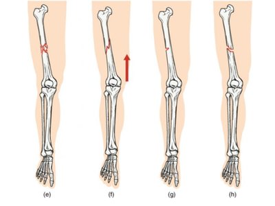
Try solving on your own before revealing the answer!
Final Answer:
(a) Transverse fracture
(b) Oblique fracture
(c) Spiral fracture
(d) Comminuted fracture
(e) Greenstick fracture
(f) Impacted fracture
(g) Segmental fracture
(h) Hairline fracture
Correct classification is important for treatment and healing.
Q12. Match the bone diseases at left with their descriptions at right.
Background
Topic: Bone Diseases
This question tests your ability to recognize and describe common bone diseases and their effects on bone structure and function.
Key Terms:
Osteomalacia
Achondroplasia
Gigantism
Osteitis Deformans
Osteopenia
Osteoporosis
Step-by-Step Guidance
Review the definitions and symptoms of each disease.
Match each disease to its description based on your knowledge.
Think about the underlying causes and effects on bone tissue.
Try solving on your own before revealing the answer!
Final Answer:
Osteomalacia: Poor bone mineralization due to inadequate Ca, PO4, or Vitamin D.
Achondroplasia: Cartilage inappropriately converted to bone, halting growth.
Gigantism: Endocrine-related growth disorder.
Osteitis Deformans: Causes deformed bone as a result of rapid reabsorption and hasty deposition of new bone matrix.
Osteopenia: Reduced bone matrix compromises bone function.
Osteoporosis: Normal age-related loss of bone.
Understanding these diseases is important for clinical practice.
Q13. What is bone remodeling? How does exercise or inactivity affect bone size? Explain Wolff’s Law.
Background
Topic: Bone Remodeling and Wolff's Law
This question tests your understanding of the dynamic nature of bone tissue and how mechanical forces influence bone structure.
Key Terms:
Bone Remodeling
Wolff's Law
Mechanical Stress
Step-by-Step Guidance
Define bone remodeling and its purpose in the body.
Explain how exercise increases bone size and strength, while inactivity leads to bone loss.
Describe Wolff’s Law and its implications for bone adaptation.
Consider why plaster casts are rarely used now and review the case study about Kevin Ware.
Try solving on your own before revealing the answer!
Final Answer:
Bone Remodeling: Continuous process of bone resorption and formation.
Exercise: Increases bone mass and strength; inactivity leads to bone loss.
Wolff’s Law: Bone adapts to the mechanical stresses placed upon it.
Plaster Casts: Less used due to risk of bone loss from inactivity.
Case Study: Kevin Ware’s injury illustrates the importance of bone remodeling in healing.
Bone health depends on mechanical stimulation and remodeling.
