 뒤로
Osseous Tissue and Bone Physiology Study Guide
스터디 가이드 - 스마트 노트
 자료에 맞춘 맞춤형 노트, 핵심 정의, 예시, 맥락을 확장해 제공합니다.
Q1. List and describe the four major functions of bone tissue:
Background
Topic: Bone Physiology
This question tests your understanding of the essential roles that bone tissue plays in the human body, which is foundational for Anatomy & Physiology.
Key Terms:
Support
Protection
Movement
Mineral Storage
Blood Cell Production
Step-by-Step Guidance
Recall that bone tissue provides structural support for the body and anchors muscles.
Think about how bones protect vital organs (e.g., skull protects the brain, ribs protect the heart and lungs).
Consider the role of bones in facilitating movement by serving as attachment points for muscles.
Remember that bones store minerals (such as calcium and phosphorus) and are involved in blood cell production (hematopoiesis) in the marrow.
Try solving on your own before revealing the answer!
Final Answer:
Support: Bones provide a framework for the body and support soft tissues.
Protection: Bones protect internal organs from injury.
Movement: Bones act as levers for muscles, enabling movement.
Mineral Storage & Blood Cell Production: Bones store minerals and house marrow for blood cell formation.
These functions are critical for maintaining homeostasis and overall health.
Q2. Complete the table describing the two types of osseous tissue:
Background
Topic: Osseous Tissue Microanatomy
This question tests your knowledge of the structural differences between compact and spongy bone, including their functional units and locations.
Key Terms:
Compact Bone (Cortical Bone)
Spongy Bone (Trabecular Bone)
Osteon
Trabeculae
Red Bone Marrow
Step-by-Step Guidance
Identify the functional unit of compact bone (hint: it's a cylindrical structure).
Recall where compact bone is located in a long bone (think about the shaft vs. ends).
Identify the functional unit of spongy bone (hint: it's a lattice-like structure).
Consider where spongy bone is found within a long bone and what it contains.

Try solving on your own before revealing the answer!
Final Answer:
Compact Bone: Functional unit is the osteon; located in the diaphysis (shaft) of long bones; provides strength and support.
Spongy Bone: Functional unit is the trabeculae; located in the epiphyses (ends) of long bones; contains red bone marrow for blood cell production.
The diagram above shows the locations of compact and spongy bone in a long bone.
Q3. Fill out the table below describing the locations and functions of these tissues: Ligament, Tendon, Articular Cartilage, Periosteum
Background
Topic: Associated Tissues of Bone Organs
This question tests your understanding of the connective tissues associated with bones and their roles in movement and protection.
Key Terms:
Ligament: Connects bone to bone
Tendon: Connects muscle to bone
Articular Cartilage: Covers joint surfaces
Periosteum: Membrane covering bone
Step-by-Step Guidance
Recall the function of ligaments and where they are found (joints).
Think about tendons and their role in transmitting muscle force to bones.
Consider the purpose of articular cartilage in joints.
Review the periosteum and its role in bone health and repair.
Try solving on your own before revealing the answer!
Final Answer:
Ligament: Connects bone to bone; stabilizes joints.
Tendon: Connects muscle to bone; transmits force for movement.
Articular Cartilage: Covers joint surfaces; reduces friction and absorbs shock.
Periosteum: Covers bone; contains nerves and blood vessels; aids in growth and repair.
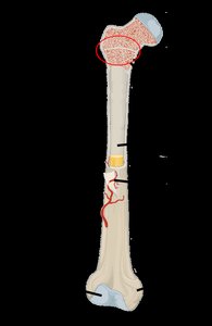
Q4. Label the indicated part of a long bone and be able to do so on an image for the exam.
Background
Topic: Macro Anatomy of Long Bones
This question tests your ability to identify and label the anatomical regions of a long bone, which is essential for understanding bone structure and function.
Key Terms:
Diaphysis
Epiphysis
Metaphysis
Medullary Cavity
Periosteum
Endosteum
Step-by-Step Guidance
Review the main regions of a long bone: shaft (diaphysis), ends (epiphyses), and the area between (metaphysis).
Identify the medullary cavity and its contents.
Locate the periosteum and endosteum on the bone diagram.
Practice labeling these parts on a diagram.
Try solving on your own before revealing the answer!
Final Answer:
Diaphysis: Shaft of the bone
Epiphysis: Ends of the bone
Metaphysis: Region between diaphysis and epiphysis
Medullary Cavity: Central cavity containing marrow
Periosteum: Outer membrane covering bone
Endosteum: Inner lining of the medullary cavity
The diagram above can be used to practice labeling these regions.
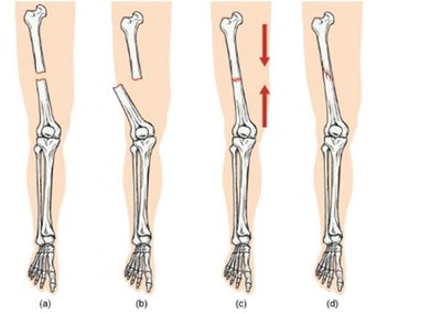
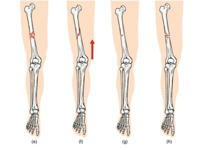
Q11. Classify the following fracture types:
Background
Topic: Bone Fractures
This question tests your ability to recognize and classify different types of bone fractures based on their appearance and characteristics.
Key Terms:
Simple (closed) fracture
Compound (open) fracture
Comminuted fracture
Greenstick fracture
Transverse fracture
Oblique fracture
Spiral fracture
Step-by-Step Guidance
Examine the images of fractured bones and note the direction and pattern of the break.
Recall the definitions of each fracture type (e.g., transverse is straight across, oblique is angled, spiral is twisted).
Match each image to the correct fracture classification based on its features.
Review the characteristics of compound vs. simple fractures.
Try solving on your own before revealing the answer!
Final Answer:
(a) Transverse fracture
(b) Oblique fracture
(c) Spiral fracture
(d) Comminuted fracture
(e) Greenstick fracture
(f) Compound (open) fracture
(g) Simple (closed) fracture
(h) Impacted fracture
The diagrams above illustrate the different fracture types for visual reference.
