 뒤로
Physiology and Anatomy of Muscle Tissue: Structure, Function, and Contraction
스터디 가이드 - 스마트 노트
 자료에 맞춘 맞춤형 노트, 핵심 정의, 예시, 맥락을 확장해 제공합니다.
Muscle Tissue: Types and Characteristics
Overview of Muscle Types
Muscle tissue is classified into three main types, each with distinct structural and functional properties:
Skeletal Muscle: Striated, voluntary, attached to bones for movement.
Cardiac Muscle: Striated, involuntary, found only in the heart.
Smooth Muscle: Non-striated, involuntary, found in walls of hollow organs.
All muscle types share common features: excitability (ability to respond to stimuli), contractility (ability to shorten), extensibility (ability to stretch), and elasticity (ability to return to original shape).
Anatomy of Skeletal Muscle
Organization and Connective Tissue Layers
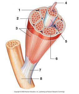
Skeletal muscle is organized into hierarchical structures, each surrounded by connective tissue layers:
Epimysium: Surrounds the entire muscle.
Perimysium: Surrounds bundles of muscle fibers called fascicles.
Endomysium: Surrounds individual muscle fibers.
Tendon: Connects muscle to bone.
Each muscle fiber contains multiple myofibrils, which are the contractile elements of the cell.
Muscle Fiber Structure
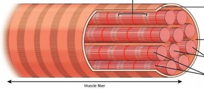
Muscle fibers (cells) are long, cylindrical, and multinucleated. They contain specialized structures:
Sarcolemma: The plasma membrane of the muscle cell.
Sarcoplasm: The cytoplasm, rich in glycosomes and myoglobin.
Myofibrils: Rod-like units composed of repeating sarcomeres.
Sarcoplasmic Reticulum (SR): Stores and releases calcium ions.
Transverse (T) Tubules: Invaginations of the sarcolemma that transmit action potentials.
Myofibrils and Sarcomere Structure
Thick and Thin Filaments
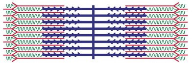
Myofibrils are composed of thick and thin filaments:
Thick Filament: Made of myosin molecules.
Thin Filament: Made of actin (G-actin and F-actin), tropomyosin, and troponin.
The arrangement of these filaments creates the striated appearance of skeletal muscle.
Sarcomere: The Functional Unit
The sarcomere is the basic contractile unit of muscle, defined by the region between two Z discs. It contains:
Z disc: Anchors thin filaments.
M line: Center of the sarcomere, anchors thick filaments.
A band: Length of thick filaments (includes overlap with thin filaments).
I band: Region with only thin filaments.
H zone: Region with only thick filaments.
Sarcoplasmic Reticulum and Triad
Structure and Function
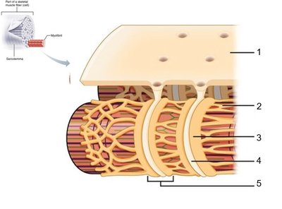
The sarcoplasmic reticulum (SR) is a specialized endoplasmic reticulum that regulates calcium ion storage and release. The triad consists of a T tubule flanked by two terminal cisternae of the SR, crucial for excitation-contraction coupling.
SR: Stores Ca2+ ions.
T tubules: Conduct action potentials deep into the muscle fiber.
Terminal cisternae: Release Ca2+ during muscle contraction.
Sliding Filament Theory and Muscle Contraction
Mechanism of Contraction
Muscle contraction occurs via the sliding filament model, where thick and thin filaments slide past each other, shortening the sarcomere:
Cross-Bridge Cycle: Myosin heads bind to actin, pull, and release, powered by ATP.
Regions: During contraction, the I band and H zone shorten, while the A band remains unchanged.
The process is regulated by Ca2+ ions and involves several steps:
Ca2+ binds to troponin, shifting tropomyosin and exposing actin binding sites.
Myosin heads attach to actin, forming cross-bridges.
ATP hydrolysis powers the movement of myosin heads.
Filaments slide, shortening the sarcomere and contracting the muscle.

Neuromuscular Junction and Excitation-Contraction Coupling
Components and Steps
The neuromuscular junction (NMJ) is the site where a motor neuron communicates with a muscle fiber:
Axon terminal: Releases acetylcholine (ACh).
Synaptic cleft: Space between neuron and muscle.
Motor end plate: Region of sarcolemma with ACh receptors.
Steps of excitation-contraction coupling:
Action potential arrives at axon terminal.
ACh is released and binds to receptors on the motor end plate.
Depolarization of sarcolemma triggers action potential in muscle fiber.
Action potential travels down T tubules, causing Ca2+ release from SR.
Ca2+ initiates contraction via the sliding filament mechanism.
Motor Units and Graded Muscle Responses
Motor Unit
A motor unit consists of a motor neuron and all the muscle fibers it innervates. The number of fibers per motor unit determines the precision of movement.
Graded Responses
Muscle contractions vary in strength depending on:
Twitch: Single, brief contraction.
Wave (temporal) summation: Increased frequency of stimulation increases contraction strength.
Unfused (incomplete) tetanus: Partial relaxation between stimuli.
Fused (complete) tetanus: No relaxation, sustained contraction.
Recruitment: Activation of additional motor units increases force.
Stimulus types:
Subthreshold stimulus: Not strong enough to cause contraction.
Threshold stimulus: Minimum stimulus required for contraction.
Maximal stimulus: Strongest stimulus, recruits all motor units.
Muscle Tone and Types of Contraction
Muscle Tone
Muscle tone is the constant, passive partial contraction of muscles, important for posture and readiness.
Types of Contraction
Isometric contraction: Muscle length does not change, tension increases.
Isotonic contraction: Muscle changes length, tension remains constant.
Concentric contraction: Muscle shortens.
Eccentric contraction: Muscle lengthens.
Energy Sources for Muscle Contraction
ATP Production Pathways
Muscle contraction requires ATP, produced by several pathways:
Creatine phosphate (CP): Rapid, short-term energy source.
Anaerobic pathway: Glycolysis produces ATP and lactic acid; used for short, intense activity.
Aerobic pathway: Uses oxygen for sustained ATP production; produces CO2 and water.
Excess Post-Exercise Oxygen Consumption (EPOC): The extra oxygen required after exercise to restore muscle to resting state.
Muscle Fiber Types
Classification and Usage
Muscle fibers are classified based on their contraction speed and metabolic properties:
Slow oxidative fibers: Fatigue-resistant, used for endurance.
Fast oxidative fibers: Intermediate properties, used for moderate activity.
Fast glycolytic fibers: Fatigue quickly, used for short bursts of power.
Smooth Muscle: Structure and Function
Comparison with Skeletal Muscle
Smooth muscle differs from skeletal muscle in several ways:
Non-striated: Lacks sarcomeres.
Involuntary: Controlled by autonomic nervous system.
Single nucleus: Per cell.
Contraction: Slower, can be sustained for longer periods.
Types of smooth muscle:
Unitary smooth muscle: Functions as a single unit, found in walls of hollow organs.
Multi-unit smooth muscle: Individual cells act independently, found in large airways and arteries.
Additional info: Academic context was added to expand brief points and ensure completeness.
