 뒤로
Regulation of Urine Concentration and Volume: Mechanisms and Clinical Evaluation
스터디 가이드 - 스마트 노트
 자료에 맞춘 맞춤형 노트, 핵심 정의, 예시, 맥락을 확장해 제공합니다.
Regulation of Urine Concentration and Volume
Osmolality and Kidney Function
The kidneys play a crucial role in maintaining the osmotic concentration of body fluids at approximately 300 mOsm. Osmolality refers to the number of solute particles in 1 kg of water, and is typically measured in milliosmols (mOsm) for biological fluids. The kidneys adjust urine concentration and volume to preserve this balance, producing concentrated urine during dehydration and dilute urine when overhydrated.
Osmolality: 1 osmol = 1 mole of particles per kg H2O; body fluids are measured in mOsm (0.001 osmol).
Urine production: Small volume when dehydrated, large volume when overhydrated.
Countercurrent Mechanisms
The kidneys utilize countercurrent mechanisms to regulate urine concentration. These mechanisms involve fluid flow in opposite directions in adjacent segments of the nephron loop and vasa recta, creating and maintaining a medullary osmotic gradient.
Countercurrent multiplier: Interaction of filtrate flow in ascending and descending limbs of nephron loops (juxtamedullary nephrons).
Countercurrent exchanger: Blood flow in ascending and descending limbs of vasa recta.
These mechanisms establish a gradient from 300 mOsm in the cortex to 1200 mOsm at the medulla's base, allowing collecting ducts to vary urine concentration.
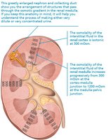
Medullary Osmotic Gradient
The medullary osmotic gradient is essential for the kidney's ability to produce urine of varying concentration. Juxtamedullary nephrons create this gradient, which is used by the collecting ducts to concentrate or dilute urine as needed.
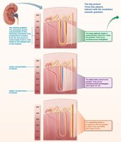
Countercurrent Multiplier
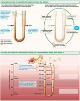
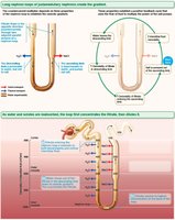
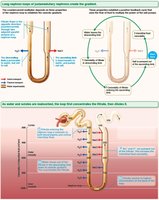
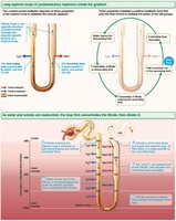
The countercurrent multiplier depends on the nephron loop's structure and function:
Filtrate flows in opposite directions in descending and ascending limbs.
Descending limb is permeable to water but not solutes; ascending limb is permeable to solutes but not water.
Active transport of NaCl out of the ascending limb increases interstitial osmolality.
A constant 200 mOsm difference exists between the limbs and between the ascending limb and interstitial fluid, multiplied along the loop's length (from 300 to 1200 mOsm).
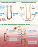
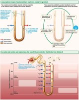
Countercurrent Exchanger
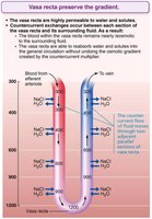
The countercurrent exchanger involves the vasa recta, which are highly permeable to water and solutes. Blood flow in the vasa recta is countercurrent, allowing exchange of NaCl and water with the interstitial fluid without disrupting the medullary gradient.
Preserves the gradient by preventing rapid removal of salt and removing reabsorbed water.
Blood remains isosmotic with interstitial fluid.
Volume of blood increases as it moves through the vasa recta.
Formation of Dilute or Concentrated Urine
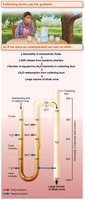
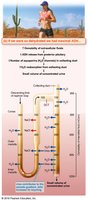
The concentration of urine depends on hydration status and the release of antidiuretic hormone (ADH):
Dehydration: Maximal ADH release, small volume of concentrated urine (~1200 mOsm).
Overhydration: Minimal ADH, large volume of dilute urine.
Urea Recycling
Urea contributes to the medullary osmotic gradient. It enters the filtrate in the ascending thin limb, is concentrated in the collecting duct, and diffuses into the medullary interstitial fluid, enhancing osmolality.
Facilitated diffusion of urea in nephron loop.
Recycling between collecting duct and ascending limb.
Clinical Evaluation of Kidney Function
Urinalysis and Renal Clearance
Urinalysis examines urine for signs of disease and illegal substances. Renal clearance measures the volume of plasma cleared of a substance per unit time, used to assess glomerular filtration rate (GFR) and detect renal disease.
Chronic renal disease: GFR < 60 ml/min for 3 months.
Renal failure: GFR < 15 ml/min, causes uremia and requires dialysis or transplant.
Urine Composition and Characteristics
Chemical Composition
95% water, 5% solutes.
Nitrogenous wastes: urea, uric acid, creatinine.
Other solutes: Na+, K+, PO43–, SO42–, Ca2+, Mg2+, HCO3–.
Abnormal components (blood proteins, WBCs, bile pigments) may indicate pathology.
Physical Characteristics
Color: Clear to deep yellow (urochrome pigment); abnormal colors may indicate disease or dietary influences.
Odor: Slightly aromatic when fresh; ammonia odor develops upon standing; altered by drugs, vegetables, or disease.
pH: Slightly acidic (~pH 6, range 4.5–8.0); affected by diet and disease.
Specific gravity: 1.001–1.035 (water = 1).
Abnormal Urinary Constituents
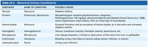
Abnormal substances in urine can indicate specific pathologies. The following table summarizes key abnormal constituents, their names, and possible causes:
Substance | Name of Condition | Possible Causes |
|---|---|---|
Glucose | Glycosuria | Diabetes mellitus |
Proteins | Proteinuria, albuminuria | Excessive physical exertion, pregnancy, hypertension, glomerulonephritis, heart failure |
Ketone bodies | Ketonuria | Starvation, diabetes mellitus |
Hemoglobin | Hemoglobinuria | Transfusion reaction, hemolytic anemia, burns |
Bile pigments | Bilirubinuria | Liver disease, obstruction of bile ducts |
Leukocytes (pus) | Pyuria | Urinary tract infection |
Clinical Homeostatic Imbalances
Renal Calculi (Kidney Stones)
Renal calculi are kidney stones formed from crystallized calcium, magnesium, or uric acid salts. Large stones can block the ureter, causing severe pain and requiring medical intervention. Risk factors include obesity and high blood calcium; prevention involves adequate hydration.
Nephrolithiasis: stones in kidneys.
Ureterolithiasis: stones in ureters.
Treatment: lithotripsy, endoscopic or surgical removal.
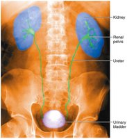
Pyelogram
A pyelogram is an imaging technique used to visualize the kidneys, renal pelvis, ureters, and bladder, aiding in the diagnosis of urinary tract disorders.
Urinary Tract Infections (UTIs)
UTIs are commonly caused by improper hygiene and anatomical factors, especially in females. Symptoms include painful urination, urgency, frequency, fever, and sometimes cloudy or blood-tinged urine. Treatment typically involves antibiotics.
Urethritis: inflammation of urethra.
Cystitis: inflammation of bladder.
Pyelitis/pyelonephritis: inflammation of kidneys.
Summary Table: Key Concepts in Urinary Regulation
Concept | Definition | Clinical Relevance |
|---|---|---|
Osmolality | Number of solute particles per kg H2O | Maintains fluid balance |
Countercurrent multiplier | Creates medullary gradient | Enables urine concentration |
Countercurrent exchanger | Preserves medullary gradient | Prevents gradient loss |
Renal clearance | Plasma volume cleared per unit time | Assesses kidney function |
Urinalysis | Examines urine for disease | Diagnostic tool |
Additional info: Academic context was added to clarify mechanisms, clinical relevance, and definitions for completeness.
