 뒤로
뒤로Sensory Physiology: Mechanisms and Pathways of Sensory Perception
스터디 가이드 - 스마트 노트
 자료에 맞춘 맞춤형 노트, 핵심 정의, 예시, 맥락을 확장해 제공합니다.
Sensory Physiology: Mechanisms and Pathways of Sensory Perception
Central Nervous System Processing of Sensory Information
The central nervous system (CNS) is responsible for receiving, integrating, and responding to sensory information. Each major division of the brain processes specific types of sensory input, allowing for appropriate responses to environmental stimuli. - Sensory Input: Sensory receptors detect stimuli and send signals to the CNS. - Integration: The CNS interprets and integrates sensory signals. - Motor Output: The CNS commands effector cells to respond, such as moving muscles. 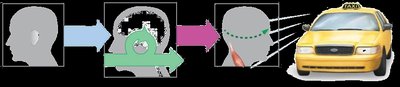
Sensory Receptors and Sensory Transduction
Sensory receptors are specialized cells that convert external or internal stimuli into electrical signals, a process known as sensory transduction. These signals are then sent to the CNS for interpretation. - Receptor Specificity: Each receptor is tuned to a specific type of stimulus (modality). - Action Potentials: All sensory receptors trigger action potentials that convey information to the CNS. - Conscious vs. Unconscious Perception: Not all sensory information results in conscious awareness; some is processed subconsciously.
Key Terminology
- Sensation: Recognition and relay of sensory stimuli to the CNS. - Perception: Conscious interpretation of sensory information. - Perception Threshold: Minimum stimulus strength required for conscious perception. - Habituation: Decreased perception due to inhibitory modulation, causing stimuli to fall below the perceptual threshold.
Types of Sensory Receptors
Sensory receptors are classified based on the type of stimulus they respond to most effectively (adequate stimulus).
Mechanoreceptors: Respond to mechanical forces (touch, pressure, vibration).
Thermoreceptors: Detect temperature changes.
Photoreceptors: Respond to light (vision).
Chemoreceptors: Detect chemical stimuli (taste, smell).
Nociceptors: Detect damaging stimuli (pain).
Sources of Sensory Input
Sensory input can originate from: - Simple Receptors: Free nerve endings (e.g., pain, smell). - Complex Receptors: Modified nerve endings (e.g., pressure, vibration). - Nonneural Receptor Cells: Specialized cells that communicate with neurons via neurotransmitters (e.g., taste, vision). 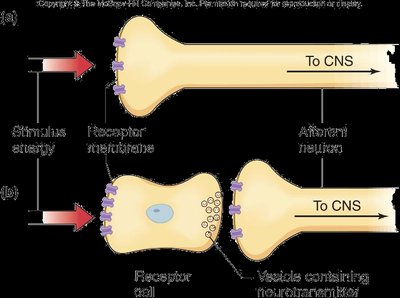
Factors Affecting Receptor Potentials
The magnitude of receptor potentials is influenced by several factors: - Stimulus Strength: Stronger stimuli produce larger receptor potentials. - Rate of Change: Gradual or abrupt changes in stimulus strength affect receptor response. - Temporal Summation: Rapid activation of receptors can sum potentials. - Adaptation: Receptors may adapt slowly (tonic) or quickly (phasic) to constant stimuli.
Receptor Adaptation: Tonic vs. Phasic Receptors
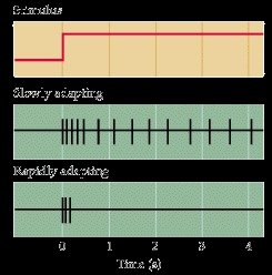
Adaptation refers to the decrease in receptor potential over time in response to a constant stimulus. - Tonic Receptors: Slowly adapting; maintain response for the duration of the stimulus (e.g., pressure, proprioception). - Phasic Receptors: Rapidly adapting; respond quickly but stop signaling under constant stimulation (e.g., smell, temperature). 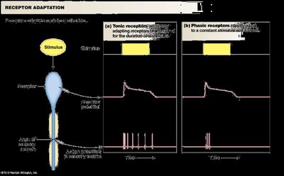
Sensory Coding: Modality, Intensity, Duration, and Location
Sensory coding is the process by which stimulus energy is converted into signals that convey relevant information to the CNS.
Modality
- Labeled Line Coding: Each receptor is associated with a specific sensation, and its neural pathway transmits that modality to the CNS.
Intensity and Duration
- Intensity: Determined by the number of receptors activated and the frequency of action potentials. - Duration: Determined by the length of receptor potentials and action potential trains. 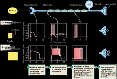
Population Coding
- Spatial Summation: Increasing the number of fibers stimulated enhances signal intensity. - Temporal Summation: Increasing the firing rate in a given number of fibers also increases intensity. 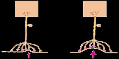
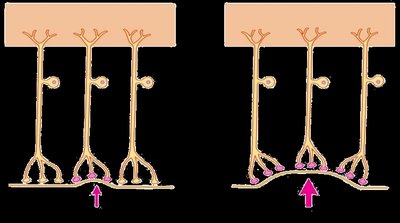
Location
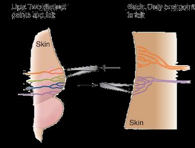
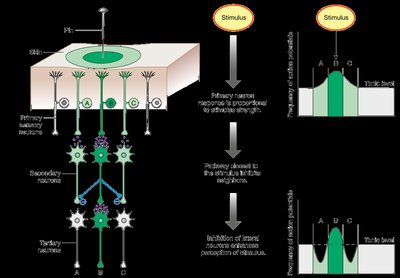
- Receptive Fields: The area within which a stimulus can activate a sensory neuron. - Acuity: The ability to discern the location of a stimulus depends on the size and overlap of receptive fields. - Lateral Inhibition: Strongly activated neurons inhibit adjacent units, enhancing localization and contrast. 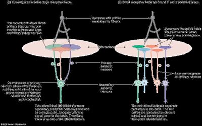
Somatic Senses: Skin and Deep Body Sensations
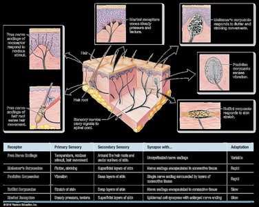
Somatic senses include touch, pressure, vibration, temperature, and pain, detected by various receptors in the skin and deeper tissues.
Free Nerve Endings: Detect temperature, noxious stimuli, and hair movement (tonic/phasic).
Meissner's Corpuscles: Detect fluttering and stroking movements (phasic).
Pacinian Corpuscles: Detect vibration (phasic).
Ruffini Corpuscles: Detect stretch (tonic).
Merkel Receptors: Detect steady pressure and texture (tonic, nonneural).
Somatosensory Pathways
Sensory information is transmitted through a series of neurons: - Primary Neuron: Projects to spinal cord or medulla. - Secondary Neuron: Decussates (crosses) and projects to the thalamus. - Tertiary Neuron: Projects to the somatosensory cortex for processing. 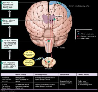
Classes of Somatosensory Nerve Fibers
Fiber Type | Characteristics | Speed of Conduction | Associated With |
|---|---|---|---|
Aβ (beta) | Large, myelinated | 30-70 m/s | Mechanical stimuli |
Aδ (delta) | Small, myelinated | 12-30 m/s | Cold, fast pain, mechanical stimuli |
C | Small, unmyelinated | 0.5-2 m/s | Slow pain, heat, cold, mechanical stimuli, itch |
Nociception: Pain Perception
Nociceptors detect damaging stimuli and are found in skin, joints, muscles, bones, and viscera. - Neurotransmitters: Substance P and glutamate. - Fiber Types: Aδ fibers convey fast, sharp pain; C fibers convey slow, dull pain. - Temperature: TRPV1 activated by heat/capsaicin; TRPM8 by cold/menthol. - Inflammatory Pain: Sensitized by inflammation and ischemia.
Pain Modulation
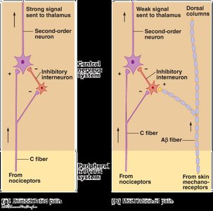
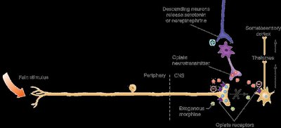
Pain perception can be modulated by various mechanisms: - Past Experiences: Learned responses can alter pain perception. - Inhibitory Interneurons: Tonically active; C fibers inhibit these, increasing pain. - Gate Control Theory: Aβ fiber stimulation excites inhibitory interneurons, reducing pain signals. - Endogenous Opioids: Endorphins, enkephalins, and dynorphins decrease neurotransmitter release and inhibit pain pathways. - Hyperalgesia: Increased sensitivity to pain, often after injury. - Pathological Pain: Chronic pain due to neurological changes. 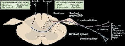
Pain Management
Analgesia is the selective suppression of pain without affecting consciousness or other sensations. - Pharmacological Agents: NSAIDs (e.g., Tylenol), opioids (e.g., morphine). - Endogenous Opioids: Released by neurons in inhibitory pathways. - Acupuncture: May activate endogenous opioid pathways. - Electrical Stimulation: TENS and CNS stimulation can reduce pain.
Referred Pain (Visceral Pain)
Referred pain is the sensation of pain at a site other than the injured tissue, often due to convergence of skin and visceral signals on second-order neurons in the spinal cord. - Organs: Few pain fibers; localized damage causes little pain, widespread damage causes severe pain. - Common Causes: Ischemia, chemical assaults, spasms, over-distention. - Localization: Pain from internal organs is often felt in the dermatome of embryological origin.
Dental Nociception
- Large Receptor Fields: Afferent fibers innervate multiple teeth, resulting in poor localization. - Dentinal Pain: Sharp pain, primarily Aδ fibers. - Pulp Pain: Dull pain, primarily C fibers. - Barrier: Dentin and pulp are separated by tight junctions, forming a selectively permeable barrier.
Summary Table: Somatic Sensory Receptors
Receptor | Primary Sensory | Secondary Sensory | Synapse With | Adaptation |
|---|---|---|---|---|
Free Nerve Endings | Temperature, noxious stimuli, hair movement | Around hair roots, under skin surface | Interneurons near endings | Variable |
Meissner's Corpuscles | Flutter, stroking | Superficial layers of skin | Interneurons near endings | Rapid |
Pacinian Corpuscles | Vibration | Deep layers of skin | Interneurons near endings | Rapid |
Ruffini Corpuscles | Stretch of skin | Deep layers of skin | Interneurons near endings | Slow |
Merkel Receptors | Steady pressure, texture | Superficial skin layers | Specialized cell synapses with enlarged nerve ending | Slow |
Summary Table: Somatosensory Pathways
Primary Sensory | Secondary Sensory | Synapse With | Tertiary Sensory |
|---|---|---|---|
Fine touch, vibration, proprioception | Medulla | Secondary sensory neuron in medulla | Somatosensory cortex |
Pain, temperature, coarse touch | Spinal cord | Secondary sensory neuron in spinal cord | Somatosensory cortex |
Key Equations
Relationship between stimulus and receptor potential:
Population Coding:
Additional info:
Some context and explanations were expanded for clarity and completeness, including definitions, examples, and summary tables to ensure the notes are self-contained and suitable for exam preparation.
