 뒤로
Skeletal Articulations and Joint Classification: Study Notes for Anatomy & Physiology
스터디 가이드 - 스마트 노트
 자료에 맞춘 맞춤형 노트, 핵심 정의, 예시, 맥락을 확장해 제공합니다.
Skeletal Articulations
Definition and Overview
Articulations, also known as arthroses, are points of contact between two bones, between bone and cartilage, or between bone and teeth. Joints are named based on the bones involved, such as the radioulnar, atlanto-occipital, glenohumeral, and acromion-clavicular (AC) joints.
Articulation: The physical connection between skeletal elements.
Joint Naming: Derived from the bones forming the joint.
Examples: Radioulnar joint, Atlanto-occipital joint, Glenohumeral joint, AC joint.
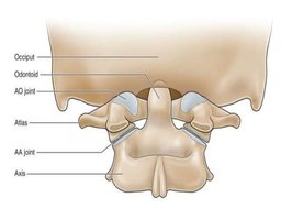
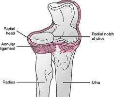
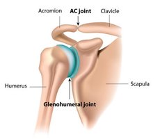
Joint Classification
Functional Classification
Joints are classified by their degree of movement:
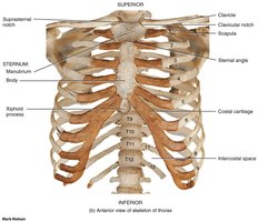
Synarthrosis: Immobile or nearly immobile joints (e.g., suture joints, cartilaginous manubriosternal joint).
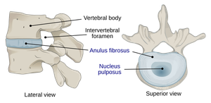
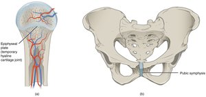
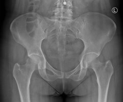
Amphiarthrosis: Limited mobility (e.g., cartilaginous joints of vertebral bodies, pubic symphysis).
Diarthrosis: Freely movable joints (all synovial joints; uniaxial, biaxial, multiaxial).
Structural Classification
Joints are also classified by the type of connective tissue and the presence or absence of a synovial cavity:
Fibrous Joints: Sutures, syndesmoses, gomphosis.
Cartilaginous Joints: Synchondroses, symphyses, epiphyseal cartilages.
Synovial Joints: Planar/gliding, saddle, pivot, hinge, condylar, ball and socket.
Fibrous Joints
General Features
Fibrous joints are held together by dense irregular connective tissue and lack a synovial cavity. They allow little to no movement.
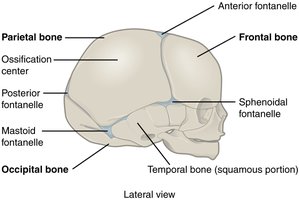
Sutures: Immobile joints found in the skull.
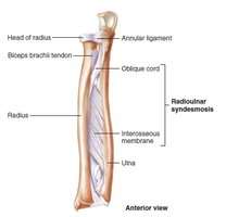
Syndesmosis: Bones connected by a sheet of fibrous tissue (interosseous membrane); allows limited movement.
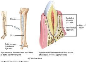
Gomphosis: Specialized joint anchoring a tooth into its socket.
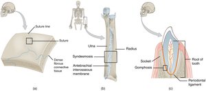
Sutures
Sutures are synarthroses in adults, providing immobility and stability to the skull.
Syndesmoses
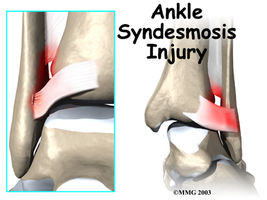
Syndesmoses have a larger distance between articulating surfaces and are connected by an interosseous membrane. They are amphiarthroses, allowing limited movement.
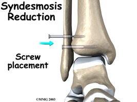
Gomphosis
Gomphosis is a synarthrosis, anchoring the root of a tooth into its socket via periodontal ligaments.
Cartilaginous Joints
General Features
Cartilaginous joints are tightly connected by hyaline cartilage or fibrocartilage and lack a synovial cavity. They allow little to no movement.
Synchondroses: Bones bound by hyaline cartilage; can be temporary (epiphyseal plate) or permanent (costochondral joints).
Symphyses: Ends of bones covered with hyaline cartilage, connected by a flat disc of fibrocartilage.
Synovial Joints
General Features
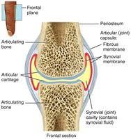
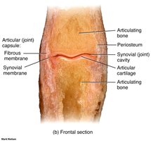
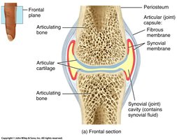
All synovial joints are diarthroses and are composed of an articular capsule, synovial cavity, synovial fluid, and articular cartilage. Accessory structures such as ligaments, discs, menisci, labrum, bursae, and tendon sheaths provide additional support.
Articular Capsule: Dense irregular connective tissue attaches to the periosteum and synovial membrane, permitting movement and preventing dislocation.
Synovial Fluid: Secreted by synovial cells; lubricates, absorbs shocks, supplies nutrients, and removes wastes.
Articular Cartilage: Hyaline cartilage lines the surfaces of articulating bones, reducing friction and absorbing shock.
Accessory Structures
Articular Disc (Meniscus): Fibrocartilage discs improve fit between condyles and circulate synovial fluid.
Labrum: Fibrocartilage rim that deepens the joint socket.
Ligaments: Extracapsular (FCL, TCL) and intracapsular (ACL, MCL) ligaments stabilize joints.
Bursae and Tendon Sheaths: Reduce friction between moving structures.
Joint Stability
Factors Affecting Stability
Articular Surfaces: Greater contact increases stability.
Ligaments: More ligaments provide greater strength and stability.
Muscle Tone: Muscle tendons crossing the joint are the most important stabilizing factor.
Movements Allowed by Synovial Joints
Types of Movement
Nonaxial: Gliding movements.
Uniaxial: Movement in one plane.
Biaxial: Movement in two planes.
Multiaxial: Movement in or around all three planes of space.
Angular Movements: Increase or decrease the angle between two bones.
Special Movements: Unique to certain joints (e.g., opposition, elevation, depression).
Types of Synovial Joints
Classification by Movement
Plane (biaxial): Gliding movements.
Hinge (uniaxial): Flexion and extension.
Pivot (uniaxial): Rotation.
Condyloid (biaxial): Flexion, extension, abduction, adduction.
Saddle (biaxial): Similar to condyloid but with greater movement.
Ball and Socket (triaxial): Movement in all axes and planes.
Summary Table: Joint Classification
Type | Structural Classification | Functional Classification | Example |
|---|---|---|---|
Fibrous | Suture, Syndesmosis, Gomphosis | Synarthrosis, Amphiarthrosis | Skull sutures, Tibiofibular syndesmosis, Tooth socket |
Cartilaginous | Synchondrosis, Symphysis | Synarthrosis, Amphiarthrosis | Epiphyseal plate, Pubic symphysis |
Synovial | Plane, Hinge, Pivot, Condyloid, Saddle, Ball & Socket | Diarthrosis | Knee, Shoulder, Hip |
Key Terms and Definitions
Articulation (Arthrosis): Point of contact between bones or between bone and cartilage/teeth.
Synarthrosis: Immobile joint.
Amphiarthrosis: Slightly movable joint.
Diarthrosis: Freely movable joint.
Synovial Fluid: Lubricating fluid in synovial joints.
Meniscus: Fibrocartilage disc in the knee.
Labrum: Fibrocartilage rim in shoulder and hip joints.
Bursa: Fluid-filled sac reducing friction.
Tendon Sheath: Tubular bursa surrounding a tendon.
Equations and Formulas
There are no specific equations for joint classification, but biomechanics may use formulas for joint forces and movement:
Joint Force: (Force equals mass times acceleration)
Torque: (Torque equals radius times force)
Examples and Applications
Clinical Example: ACL tears are common in athletes due to abnormal knee positioning or rapid direction changes.
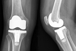
Medical Application: Joint replacement surgery (e.g., knee or hip) restores function in damaged synovial joints.
Additional info: The notes have been expanded with academic context to ensure completeness and clarity for exam preparation.
