 뒤로
Skeletal Muscle Contraction and Energy Sources: Study Guide
스터디 가이드 - 스마트 노트
 자료에 맞춘 맞춤형 노트, 핵심 정의, 예시, 맥락을 확장해 제공합니다.
Skeletal Muscle Contraction
Overview of Muscle Contraction
Skeletal muscle contraction is a complex physiological process that enables voluntary movement. It involves a series of coordinated events at the cellular and molecular levels, including excitation, excitation-contraction coupling, and the contraction cycle.
Excitation: Initiated by a nerve impulse at the neuromuscular junction.
Excitation-Contraction Coupling (ECC): Links the electrical signal to muscle contraction via calcium release.
Contraction: The sliding filament mechanism produces force and movement.
Neuromuscular Junction (NMJ)
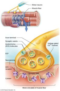
The neuromuscular junction is the synapse between a motor neuron and a skeletal muscle fiber. It is essential for transmitting the nerve impulse that triggers muscle contraction.
Motor Neuron: Releases acetylcholine (ACh) from synaptic vesicles.
Synaptic Cleft: The gap between the neuron and muscle fiber.
Motor End Plate: Region of the muscle fiber membrane with ACh receptors.
Excitation Phase
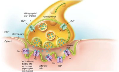
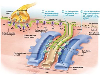
During excitation, the motor neuron releases acetylcholine into the synaptic cleft, which binds to receptors on the motor end plate, causing depolarization of the sarcolemma.
Voltage-gated Ca2+ channels: Open in the axon terminal, allowing Ca2+ influx.
ACh release: Triggers Na+ influx in the muscle fiber, generating an action potential.
Excitation-Contraction Coupling (ECC)
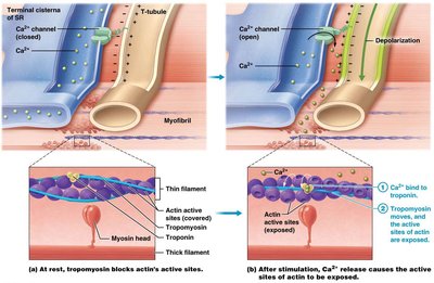
ECC is the process by which the action potential in the muscle fiber leads to the release of calcium ions from the sarcoplasmic reticulum (SR), initiating contraction.
T-tubules: Conduct the action potential deep into the muscle fiber.
Calcium Release: Ca2+ channels open, releasing Ca2+ into the sarcoplasm.
Troponin and Tropomyosin: Ca2+ binds to troponin, shifting tropomyosin and exposing actin binding sites.
Contraction Cycle (Sliding Filament Theory)
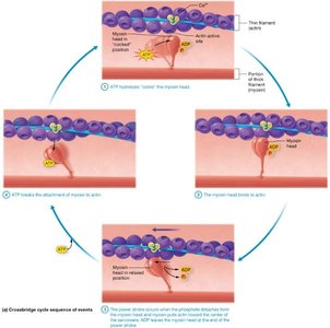
The contraction cycle describes how myosin heads bind to actin, pull, and release, resulting in muscle shortening.
Cross-Bridge Formation: Myosin heads bind to exposed actin sites.
Power Stroke: Myosin pulls actin, shortening the sarcomere.
ATP Role: ATP binds to myosin, causing detachment and re-cocking of the head.
Muscle Relaxation
Muscle relaxation occurs when stimulation ceases, and calcium is pumped back into the SR.
Acetylcholinesterase: Breaks down ACh, ending stimulation.
Return to Resting Membrane Potential: Sarcolemma repolarizes.
Calcium Removal: Ca2+ is actively transported back into the SR.
Tropomyosin: Covers actin binding sites, preventing contraction.
Clinical Applications: Neuromuscular Blockers
Botulinum Toxin (BoTox)
Source: Clostridium botulinum
Mechanism: Blocks presynaptic release of acetylcholine, causing paralysis.
Clinical Use: Used therapeutically to treat muscle spasticity and cosmetically to reduce wrinkles.
Curare
Source: Chondrodendron tomentosum
Mechanism: Binds to ACh receptors, preventing nerve impulses from activating muscles.
Clinical Use: Used as a neuromuscular blocking agent in anesthesia.
Energy Sources for Muscle Contraction
ATP Requirements
ATP is essential for muscle contraction and relaxation. It powers the Na+/K+ pumps, the myosin power stroke, and the active transport of Ca2+ back into the SR.
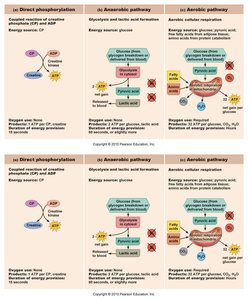
Immediate ATP Source: Creatine Phosphate
Creatine phosphate provides a rapid means of regenerating ATP during short bursts of activity.
Creatine Kinase: Catalyzes the transfer of a phosphate group to ADP, forming ATP.
Storage: Skeletal muscles have a maximal storage capacity for creatine.
Anaerobic Cellular Respiration: Glycolytic Energy Sources
Anaerobic glycolysis produces ATP without oxygen, but results in lactic acid buildup.
Requirements: Glucose
Products: 2 ATP, lactic acid
Lactic Acid: Accumulates during intense activity, causing muscle fatigue.
Aerobic Cellular Respiration: Oxidative Energy Sources
Aerobic metabolism uses oxygen to produce large amounts of ATP from glucose, fatty acids, and amino acids.
Requirements: Oxygen, fuel molecules
Products: Many ATP, CO2, H2O
Sources of Oxygen: Blood and myoglobin in muscle fibers
Excess Post-exercise Oxygen Consumption (EPOC)
EPOC refers to the increased rate of ventilation after exercise, necessary to restore homeostasis.
Heat Dissipation: Removes excess heat generated during activity.
Restoration of Ion Concentrations: Rebalances Na+, K+, and Ca2+ levels.
Correction of Blood pH: Eliminates lactic acid and restores normal pH.
Summary Table: Muscle Energy Pathways
Pathway | Requirements | Products | Duration |
|---|---|---|---|
Direct Phosphorylation (Creatine Phosphate) | CP, ADP | ATP, Creatine | Short (seconds) |
Anaerobic Glycolysis | Glucose | ATP, Lactic Acid | Short (minutes) |
Aerobic Respiration | Glucose, O2 | ATP, CO2, H2O | Long (hours) |
Key Terms and Definitions
Excitation-Contraction Coupling: The process linking muscle fiber excitation to contraction.
Neuromuscular Junction: Synapse between motor neuron and muscle fiber.
Acetylcholine: Neurotransmitter released at NMJ.
Creatine Phosphate: High-energy compound used for rapid ATP regeneration.
Glycolysis: Anaerobic breakdown of glucose.
Oxidative Catabolism: Aerobic breakdown of fuel molecules.
Relevant Equations
ATP regeneration from creatine phosphate: $\text{CP} + \text{ADP} \rightarrow \text{Creatine} + \text{ATP}$
Anaerobic glycolysis: $\text{Glucose} \rightarrow 2 \text{ATP} + \text{Lactic Acid}$
Aerobic respiration: $\text{Glucose} + 6 \text{O}_2 \rightarrow 36 \text{ATP} + 6 \text{CO}_2 + 6 \text{H}_2\text{O}$
Example Application
Creatine Supplementation: May improve performance in short, intense activities but can cause water retention and kidney damage if taken in excess.
Botulinum Toxin: Used clinically to treat muscle spasticity and cosmetically to reduce wrinkles by causing temporary muscle paralysis.
Additional info: Expanded explanations and context were added to ensure completeness and academic quality.
