 뒤로
Skeletal Muscle Contraction: Structure, Function, and Mechanisms
스터디 가이드 - 스마트 노트
 자료에 맞춘 맞춤형 노트, 핵심 정의, 예시, 맥락을 확장해 제공합니다.
Muscles and Muscle Tissue
Microscopic Structure and Functional Roles of Skeletal Muscle Fibers
Skeletal muscle fibers are highly specialized cells responsible for voluntary movement. Their structure is essential for their function, and several key components play distinct roles in muscle contraction.
Myofibrils: Cylindrical structures within muscle fibers composed of repeating units called sarcomeres, which are the functional contractile units. Myofibrils contain thick (myosin) and thin (actin) filaments.
Sarcoplasmic Reticulum (SR): A specialized endoplasmic reticulum that stores and releases calcium ions (Ca2+), which are crucial for initiating contraction.
T Tubules: Invaginations of the sarcolemma (muscle cell membrane) that transmit action potentials deep into the muscle fiber, ensuring coordinated contraction.
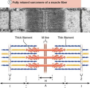
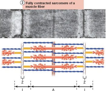
Sarcomere: The basic contractile unit, defined by Z discs, containing overlapping thick and thin filaments.
Example: The sarcomere shortens during contraction, leading to muscle fiber shortening and movement.
Sliding Filament Model of Muscle Contraction
The sliding filament model explains how muscles contract at the molecular level. It describes the interaction between actin and myosin filaments within the sarcomere.
Contraction: Activation of cross bridges (myosin heads binding to actin) generates force.
Shortening: Occurs when tension generated by cross bridges exceeds opposing forces, pulling thin filaments toward the center of the sarcomere.
Filament Overlap: During contraction, thin filaments slide past thick filaments, increasing overlap. Neither filament changes length.
Cross Bridge Cycling: Myosin heads attach, pivot, detach, and re-cock, pulling actin filaments in a ratcheting action.
Structural Changes: I bands shorten, Z discs move closer, H zones disappear, and A bands move closer together.
Example: Muscle contraction is initiated when the nervous system stimulates the muscle fiber, allowing myosin heads to bind to actin.
Motor Neurons Stimulate Skeletal Muscle Fibers to Contract
Neuromuscular Junction and Muscle Fiber Stimulation
The neuromuscular junction (NMJ) is the site where motor neurons communicate with muscle fibers to initiate contraction. This process involves several steps:
Motor Neurons: Somatic motor neurons transmit signals from the brain and spinal cord to skeletal muscles.
Axon Terminals: Each axon divides into branches ending at the muscle fiber, forming the NMJ.
Synaptic Cleft: The space between the axon terminal and muscle fiber, filled with gel-like material.
Synaptic Vesicles: Contain the neurotransmitter acetylcholine (ACh), released upon stimulation.
Junctional Folds: Infoldings of the sarcolemma containing ACh receptors.
Example: Each muscle fiber has one NMJ with one motor neuron.
Events at the Neuromuscular Junction
Muscle contraction begins with a series of events at the NMJ:
Action potential arrives at the axon terminal.
Voltage-gated calcium channels open; Ca2+ enters the motor neuron.
Ca2+ entry triggers release of ACh into the synaptic cleft.
ACh diffuses across the cleft and binds to ACh receptors on the sarcolemma.
ACh binding opens Na+ channels, resulting in an end plate potential (local depolarization).
Acetylcholinesterase degrades ACh, terminating the signal.
Generation and Propagation of Action Potential Across the Sarcolemma
The action potential (AP) is a rapid change in membrane voltage that triggers muscle contraction. The process involves:
End Plate Potential: Local depolarization caused by Na+ influx after ACh binds to its receptor.
Depolarization: If threshold is reached, voltage-gated Na+ channels open, causing a large influx of Na+ and propagation of the AP.
Repolarization: Na+ channels close, K+ channels open, restoring resting membrane potential. The Na+-K+ pump reestablishes ionic conditions.
Refractory Period: The muscle fiber cannot be stimulated again until repolarization is complete.
Example: The AP spreads across the sarcolemma, leading to muscle fiber contraction.
Excitation-Contraction Coupling
Linking Excitation to Contraction
Excitation-contraction (E-C) coupling refers to the events that link the AP on the sarcolemma to the sliding of myofilaments and muscle contraction.
AP Propagation: The AP travels along the sarcolemma and into T tubules.
Ca2+ Release: Voltage-sensitive proteins in T tubules stimulate Ca2+ release from the SR.
Contraction Initiation: Ca2+ binds to troponin, causing tropomyosin to move and exposing myosin-binding sites on actin.
Cross Bridge Formation: Myosin heads bind to actin, initiating contraction.
Example: E-C coupling ensures that electrical signals result in mechanical contraction.
Cross Bridge Cycle
The cross bridge cycle is the sequence of events that leads to muscle contraction:
Cross Bridge Formation: High-energy myosin head attaches to actin.
Power Stroke: Myosin head pivots, pulling actin toward the M line.
Cross Bridge Detachment: ATP binds to myosin, causing it to detach from actin.
Cocking of Myosin Head: ATP hydrolysis re-energizes the myosin head for the next cycle.
Example: The steps of the cross bridge cycle are analogous to pulling a rope with one hand.
Clinical Relevance: Homeostatic Imbalances
Myasthenia Gravis
Myasthenia gravis is an autoimmune disease where antibodies attack ACh receptors, leading to muscle weakness, drooping eyelids, and difficulty swallowing and talking.
Mechanism: Fewer ACh receptors reduce muscle fiber stimulation.
Symptoms: Generalized muscle weakness and fatigue.
Rigor Mortis
Rigor mortis is the stiffening of muscles after death, typically beginning 3–4 hours postmortem and peaking at 12 hours.
Cause: ATP production ceases, Ca2+ cannot be pumped back into the SR, resulting in sustained cross bridge formation.
Detachment Failure: ATP is required for cross bridge detachment; without it, myosin remains bound to actin.
Resolution: Muscles remain contracted until proteins break down, releasing myosin.
Summary Table: Steps of Skeletal Muscle Contraction
Step | Description |
|---|---|
1. Events at NMJ | AP arrives, ACh released, binds to receptors, Na+ influx |
2. Muscle Fiber Excitation | End plate potential, depolarization, AP propagation |
3. Excitation-Contraction Coupling | AP triggers Ca2+ release, troponin activation |
4. Cross Bridge Cycling | Myosin binds actin, power stroke, detachment, re-cocking |
Key Equations
Resting Membrane Potential:
Na+-K+ Pump:
Additional info: Academic context was added to clarify the structure and function of muscle fibers, the sliding filament model, and clinical relevance. Images were included only when directly relevant to the explanation of sarcomere structure and neuromuscular junction anatomy.
