 뒤로
Skeletal Muscle Structure and Function: Study Notes for Anatomy & Physiology L12
스터디 가이드 - 스마트 노트
 자료에 맞춘 맞춤형 노트, 핵심 정의, 예시, 맥락을 확장해 제공합니다.
Skeletal Muscle: Overview and Types
Muscle Tissue Types
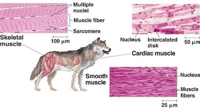
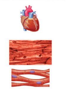
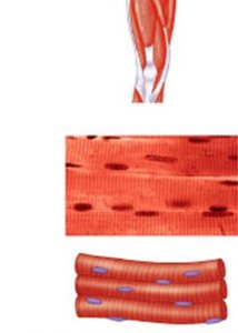
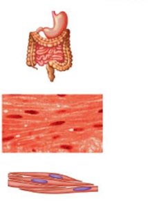
Muscle tissue is specialized for excitability and contraction, enabling movement throughout the body. There are three main types of muscle tissue, each with distinct structural and functional characteristics:
Skeletal Muscle: Moves the skeleton, is striated, voluntary, and contains multiple nuclei per cell.
Cardiac Muscle: Found in the heart, is striated, involuntary, and typically has one nucleus per cell.
Smooth Muscle: Located in hollow organs, is non-striated, involuntary, and has one nucleus per cell.
Skeletal Muscle Organization
Muscle Structure: Organ to Cell
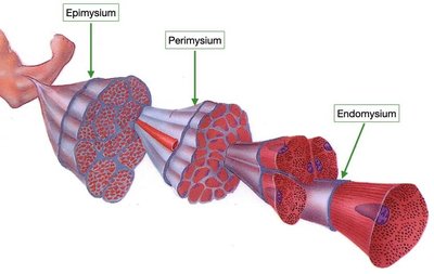
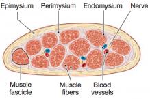
Skeletal muscle is organized hierarchically, from the whole muscle organ down to individual muscle fibers (cells). Each level is surrounded by connective tissue layers that provide support and facilitate force transmission.
Epimysium: Dense irregular connective tissue surrounding the entire muscle.
Perimysium: Dense irregular connective tissue surrounding bundles of muscle fibers called fascicles.
Endomysium: Connective tissue surrounding each individual muscle fiber.
Connective Tissue Attachments
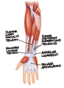
Connective tissue layers are continuous with tendons and aponeuroses, which anchor muscles to bones and facilitate movement.
Tendons: Bundles of dense regular connective tissue that attach muscle to bone.
Aponeuroses: Broad, flat sheets of tendon that cover muscles and attach to bone.
Skeletal Muscle Function
General and Specialized Functions
Skeletal muscle serves multiple roles in the body, including movement, posture, and protection. Specialized functions include:
Guarding entrances and exits of the body (e.g., sphincters)
Maintaining body temperature (e.g., shivering)
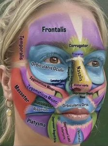
Emotional expression (e.g., facial muscles)
Ventilation (e.g., diaphragm changes lung volume)
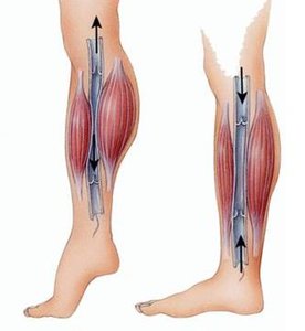
Assisting blood flow, especially in lower limbs
Fascicle Arrangement and Muscle Power
Fascicle Arrangements
The arrangement of fascicles within a muscle determines its power and range of motion. There are four major types:
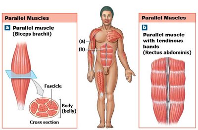
Parallel: Fascicles run parallel to the long axis; efficient for fast, smooth movement. Examples: biceps brachii, rectus abdominus.
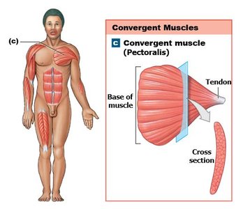
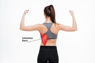
Convergent: Fascicles converge from a broad area to a single point; more fibers generate more force. Examples: pectoralis major, latissimus dorsi.
Circular: Fascicles form rings around openings; contract to close and relax to open. Examples: orbicularis oculi, orbicularis oris.
Pennate: Fascicles attach obliquely to a common tendon; more fibers generate more force, but shorter length reduces range of motion. Types: unipennate (extensor digitorum), bipennate (rectus femoris), multipennate (deltoid).
Skeletal Muscle Fiber Structure
Muscle Fiber Components
Each skeletal muscle fiber is a single, elongated cell containing specialized structures for contraction:
Sarcolemma: Cell membrane of the muscle fiber.
Transverse (T) Tubules: Infoldings of the sarcolemma that help transmit action potentials.
Sarcoplasmic Reticulum: Specialized smooth endoplasmic reticulum that stores calcium ions (Ca2+).
Terminal Cisternae: Enlarged ends of the sarcoplasmic reticulum.
Myofibrils: Chains of sarcomeres, the functional contractile units.
Sarcomere Structure
The sarcomere is the basic contractile unit of muscle, defined by the region between two Z discs. It contains:
Z Disc: Boundary of each sarcomere, anchors thin filaments (actin).
M Line: Center of the sarcomere, anchors thick filaments (myosin).
I Band: Region containing only thin filaments.
H Zone: Central region containing only thick filaments.
A Band: Entire length of thick filaments, including overlap with thin filaments.
Titin: Elastic protein connecting myosin to Z disc, helps restore sarcomere length after contraction.
Sliding Filament Theory of Contraction
Mechanism of Muscle Contraction
Muscle contraction occurs when sarcomeres shorten due to the sliding of actin (thin) and myosin (thick) filaments past each other. This process is powered by ATP and involves several steps:
Cross-Bridge Formation: Myosin head attaches to actin.
Power Stroke: Myosin head pivots, pulling actin toward the center of the sarcomere.
Cross-Bridge Detachment: ATP binds to myosin, causing it to release actin.
Reactivation: ATP is hydrolyzed, re-cocking the myosin head for another cycle.
The cycle continues as long as ATP is available and calcium ion concentration in the sarcoplasm remains high.
Key Equations
The energy for muscle contraction is provided by ATP hydrolysis:
Force generation depends on the number of cross-bridges formed:
Summary Table: Muscle Types
Type | Location | Striations | Control | Number of Nuclei |
|---|---|---|---|---|
Skeletal | Bones | Yes | Voluntary | Many |
Cardiac | Heart | Yes | Involuntary | One |
Smooth | Hollow Organs | No | Involuntary | One |
Additional info:
These notes expand on the original lecture slides and questions, providing full academic context and definitions for all key terms. The included images directly reinforce the structural and functional concepts of muscle tissue, organization, and contraction mechanisms.
