 뒤로
Skeletal Muscle Structure and Function: Study Notes for Anatomy & Physiology
스터디 가이드 - 스마트 노트
 자료에 맞춘 맞춤형 노트, 핵심 정의, 예시, 맥락을 확장해 제공합니다.
Muscle Tissue: Properties and Types
General Properties of Muscle Tissue
Muscle tissue is essential for movement and force generation in the body. It possesses several unique properties that distinguish it from other tissue types:
Excitability: Ability to respond to stimuli.
Contractility: Muscle cells shorten when stimulated, producing force.
Extensibility: Ability to stretch without being damaged.
Elasticity: Ability to return to original shape after stretching or contracting.
Types of Muscle Tissue
There are three main types of muscle tissue, each with distinct characteristics:
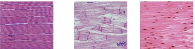
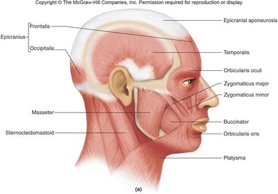
Skeletal Muscle: Striated, voluntary, multinucleate, moves bones and facial skin.
Cardiac Muscle: Striated, involuntary, contains gap junctions, coordinated activity allows it to act as a pump.
Smooth Muscle: Non-striated, involuntary, found in walls of hollow organs.
Example: Skeletal muscle contracts to move limbs, cardiac muscle contracts to pump blood, and smooth muscle contracts to move food through the digestive tract.
Skeletal Muscle Structure and Organization
Levels of Organization
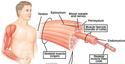
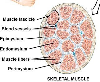
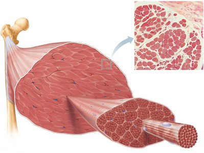
Skeletal muscle is organized in a hierarchical structure, with bundles within bundles:
Epimysium: Surrounds the entire muscle.
Perimysium: Surrounds each fascicle (bundle of muscle fibers).
Endomysium: Surrounds individual muscle fibers (cells).
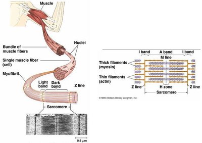
Each muscle fiber contains myofibrils, which are composed of myofilaments (actin and myosin). Blood vessels and nerves are present within each fascicle, and neurons innervate individual muscle fibers. Myosatellite stem cells help repair muscle tissue.
Muscle Fiber Anatomy
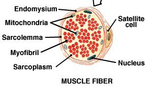
Muscle fibers are specialized cells with unique structures:
Sarcolemma: Plasma membrane of muscle cell.
Sarcoplasm: Cytoplasm of muscle cell.
Sarcoplasmic Reticulum: Specialized endoplasmic reticulum for calcium storage.
Multinucleate: Muscle fibers contain multiple nuclei.
The prefixes "myo" and "sarco" indicate muscle-related structures (e.g., myofibril, myoglobin).
Myofibrils and Sarcomeres
Myofibrils are composed of repeating units called sarcomeres, which are the functional units of muscle contraction. Each sarcomere contains:
Actin (thin) filaments
Myosin (thick) filaments
Z line: Boundary of the sarcomere
M line: Center of the sarcomere
I band: Region with only actin
A band: Region with both actin and myosin
H zone: Region with only myosin
Skeletal Muscle Attachments and Movement
Muscle Attachments
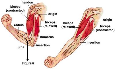
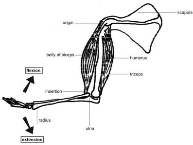
Skeletal muscles span joints and attach to bones at two main points:
Origin: The less movable attachment, usually proximal.
Insertion: The more movable attachment, usually distal.
Muscles are commonly attached to bone via tendons or aponeuroses. Muscle contraction pulls on connective tissue sheaths, transmitting force to the bone.
Muscle Fiber Formation and Development
Muscle Fiber Formation
Muscle fibers form during embryonic development (by day 45-50) and can also form due to mechanical strain. Muscle cells are multinucleate due to the fusion of myoblasts during development.
Typical body cells: Single nucleus, plasma membrane, cytoplasm, endoplasmic reticulum.
Muscle cells: Multinucleate, sarcolemma, sarcoplasm, sarcoplasmic reticulum.
Example: The prefix "myo" or "sarco" in terms like myoblast or sarcoplasmic reticulum indicates muscle tissue involvement.
Mechanism of Skeletal Muscle Contraction
Excitation-Contraction Coupling
Skeletal muscle contraction is triggered by neural stimulation at the neuromuscular junction (NMJ). The process involves:
Activation: Neural stimulation at NMJ.
Excitation-contraction coupling: Generation and propagation of an action potential along the sarcolemma.
Increase in intracellular Ca2+: Ca2+ released from the sarcoplasmic reticulum.
Neuromuscular Junction (NMJ)
The NMJ is the site where a motor neuron communicates with a muscle fiber:
Axon terminal: Releases acetylcholine (ACh) neurotransmitter.
Synaptic cleft: Gap between axon terminal and muscle fiber.
Junctional folds: Contain ACh receptors on the sarcolemma.
When ACh binds to its receptors, it triggers an action potential in the muscle fiber.
Steps in Muscle Contraction and Relaxation
The process of muscle contraction and relaxation involves several steps:
Step | Initiating Contraction | Muscle Relaxation |
|---|---|---|
1 | ACh released, binding to receptors | ACh broken down by AChE |
2 | Action potential reaches T tubule | Sarcoplasmic reticulum recaptures Ca2+ |
3 | Sarcoplasmic reticulum releases Ca2+ | Active sites covered, no cross-bridge interaction |
4 | Active site exposure, cross-bridge formation | Contraction ends |
5 | Contraction begins | Relaxation occurs, passive return to resting length |
Sliding Filament Theory
Muscle contraction occurs when myosin heads bind to actin, forming cross-bridges and pulling the actin filaments toward the M line. ATP is required for cross-bridge detachment and re-cocking of the myosin head. If Ca2+ remains bound to troponin and ATP is available, the cycle repeats.
H zone and I band shorten during contraction.
ATP hydrolysis:
Muscle Relaxation and Chemical Interference
Muscle Relaxation
Muscle relaxation occurs when neural stimulation ceases, Ca2+ is pumped back into the sarcoplasmic reticulum, and ATP is available. Elastic elements, antagonistic muscle contractions, and gravity help return muscles to resting length.
Chemical Interference
Certain chemicals can interfere with muscle contraction:
Rigor Mortis: Occurs 2-7 hours after death due to lack of ATP, preventing Ca2+ reuptake and cross-bridge detachment.
Botox: Blocks ACh release, causing local paralysis.
Curare: Inhibits ACh receptors, leading to paralysis.
ACh esterase antagonists: Prevent breakdown of ACh, causing prolonged contraction.
Example: Insecticides that inhibit ACh esterase cause continuous muscle contraction in insects.
Key Terms and Concept Mapping
Action potential
Acetylcholine
Acetylcholine esterase
Axon
Excitation-contraction coupling
Ligand-gated channels
Motor neuron
Neuromuscular junction
Neurotransmitter
Synapse
Synaptic cleft
Voltage-gated channels
Sarcoplasmic reticulum
Sarcomere
Muscle fiber
